Diversity of Life: Domains Bacteria and Archaea with a Look at Viruses
Diversity of Life: Domains Bacteria and Archaea with a Look at Viruses
PROKARYOTES
This section considers three different groups of very different “organisms.” The first group includes those organisms that have prokaryotic cells. These are usually referred to as bacteria although it is a heterogeneous group. These organisms have several unique characteristics.
1. They have prokaryotic cells. These cells have no membrane bound organelles—in particular the nucleus but also no mitochondria, endoplasmic reticulum, Golgi apparatus, chloroplasts, etc. These cells also have a cell wall containing a uniquely prokaryotic substance called peptidoglycan. They may also have an outer membrane outside of the cell wall and/or a mucous capsule surrounding the entire cell.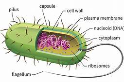
2. Prokaryotic cells are much smaller than cells of other organisms. They are about 1/10 the size of most eukaryotic cells.
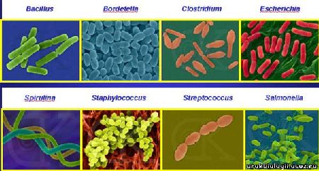
3. Most bacterial cells have one of three shapes—a bacillus (rod-shaped), a coccus (ball-shaped), or a spirochaete (corkscrew-shaped).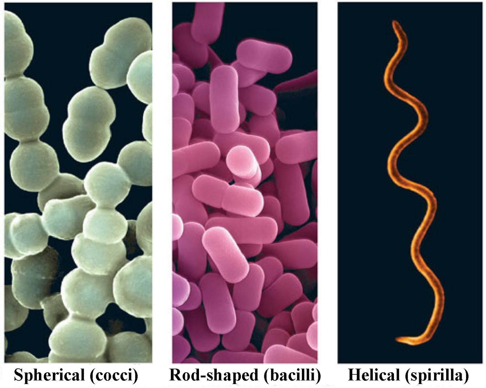
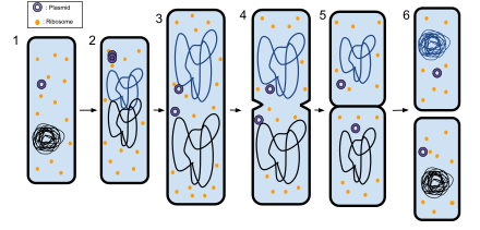
4. Prokaryotic cells do NOT divide by mitosis or meiosis. Instead they use a very different cell reproduction process called bacterial fission. Since they usually have only one, circular chromosome, the chromosome replicates and the two chromosomes attach to the cell membrane. Then, cell wall material is added between the two chromosomes, pushing them apart. When far enough apart, the cell divides into two cells. They use conjugation as a means of exchanging DNA, usually between plasmids.
5. Bacteria exhibit metabolic diversity. Most other groups of organisms have a single process for obtaining both nutrients and energy. Bacteria show four different processes.
- Photoautotrophs: Use sunlight as an energy source and inorganic carbon (usually CO2) as a carbon source.
- Photoheterotrophs: Use sunlight as an energy source and organic carbon as a carbon source.
- Chemoautilithotrophs: Oxidize inorganic compounds as an energy source and inorganic carbon (usually CO2) as a carbon source.
- Chemoorganotrophs: Use organic compounds as both an energy source and nutrient source.
6. A few bacteria form a resistant stage called an endospore during adverse conditions.
All prokaryotic organisms used to be in a single kingdom--Monera. However, as the technology to investigate the biochemistry of these tiny organisms improved, scientists realized that there were actually two very different groups of prokaryotes with very different cell wall construction and ribosome structure. In fact, the difference between the two groups was as large as the difference between either one and eukaryote cells. As a result, the classification category Domain was created with two prokaryotic Domains—Bacteria and Archaea.
Here is a PBS NOVA article on how Dr. Carl Woese figured out that the prokaryotic organisms in the Kingdom Monera belonged in two very different groups. This shows the how the power of the process of science and how advances in technology lead to new discoveries.
For a slightly more detailed look at how he did this work, look at the following video.
The Domain Bacteria contains most of the organisms we commonly refer to as bacteria, including the less than one percent that are pathogenic. This group also includes photosynthetic bacteria called cyanobacteria (formerly the blue-green algae). Many of these organisms are critical for ecosystem functioning or are used by humans beneficially.
The Archaea, on the other hand, are not pathogens and are of great interest because they are often found in “extreme” environments—high salt (halophiles), high or low temperatures (thermophiles), high acid (acidophiles), and anaerobic environments (methanogens).
 Yellowstone Park hot spring where Archaeans were first identified
There are three basic types of Archaeans, each found in a unique environment that is hostile to other forms of life: methanogens, halophiles, and thermoacidophiles. See this link on Finding Life's Extremists for more details.
VIRUSES
Viruses are noncellular—no cell membrane, no organelles, no metabolic activity on their own, unable to reproduce on their own. In short, they exhibit none of the characteristics of life that we covered in the first chapter. They are not put into any of the domains or kingdoms used to classify living organisms. They do, however, enter living host cells and “take over” those cells to make new virus particles. They consist of only two parts: an inner core of nucleic acid (either DNA or RNA and a protein capsid surrounding the nucleic acid. A few may also have a capsule surrounding the virus particle that is usually made in part from the host cell membrane.
There are two other groups that are even more strange than the bacteria and viruses.
VIROIDS
Little is really known about these infectious molecules. They consist of naked strands of RNA and infect plants.
PRIONS
Around 1900 a disease was identified among the South Fore people of New Guinea; it was called Kuru. It is a neurodegenerative disease and it was not until the 1960’s that researchers began to understand how it was transmitted. The South Fore were cannibals; not in the sense of eating strangers or enemies but their funeral rituals involved honoring the dead by eating them. Women and children contracted the disease more than men because they ate the organs while the men ate mostly the muscle.
In sheep, a disease called scrapie has long been known. In addition, another neurodegenerative disease found on rare occasions in Europe and North America was called Creutzfeldt-Jacob disease. The cause of all of these was unknown until the 1990’s. Then, Stanley Prusiner advocated a radical solution for the cause. He said it was caused by an infectious protein called a prion. There is no genetic material.
Recall that every protein has a unique three dimensional shape. The prion protein is a normal protein found in neurons. However, it can have one of two shapes. The normal shape is what is usually found in cells. However, if an abnormally shaped prion protein enters the cell it is able to slowly convert the normal proteins to the abnormal form. Eventually this conversion causes death of the cell. Eventually enough cells die to cause the characteristic spongy appearance of the brain tissue found upon autopsy. This same prion also causes bovine spongiform encephalopathy (BSE), more popularly known as mad cow disease. We now know that ¼ teaspoon of infected tissue consumed by a cow can be enough to transmit the disease.