Spinal Cord and Relexes
Spinal Cord and Relexes
Characteristics of CNS- 4 major characteristics
1. Protected by bone - skull & vertebrae
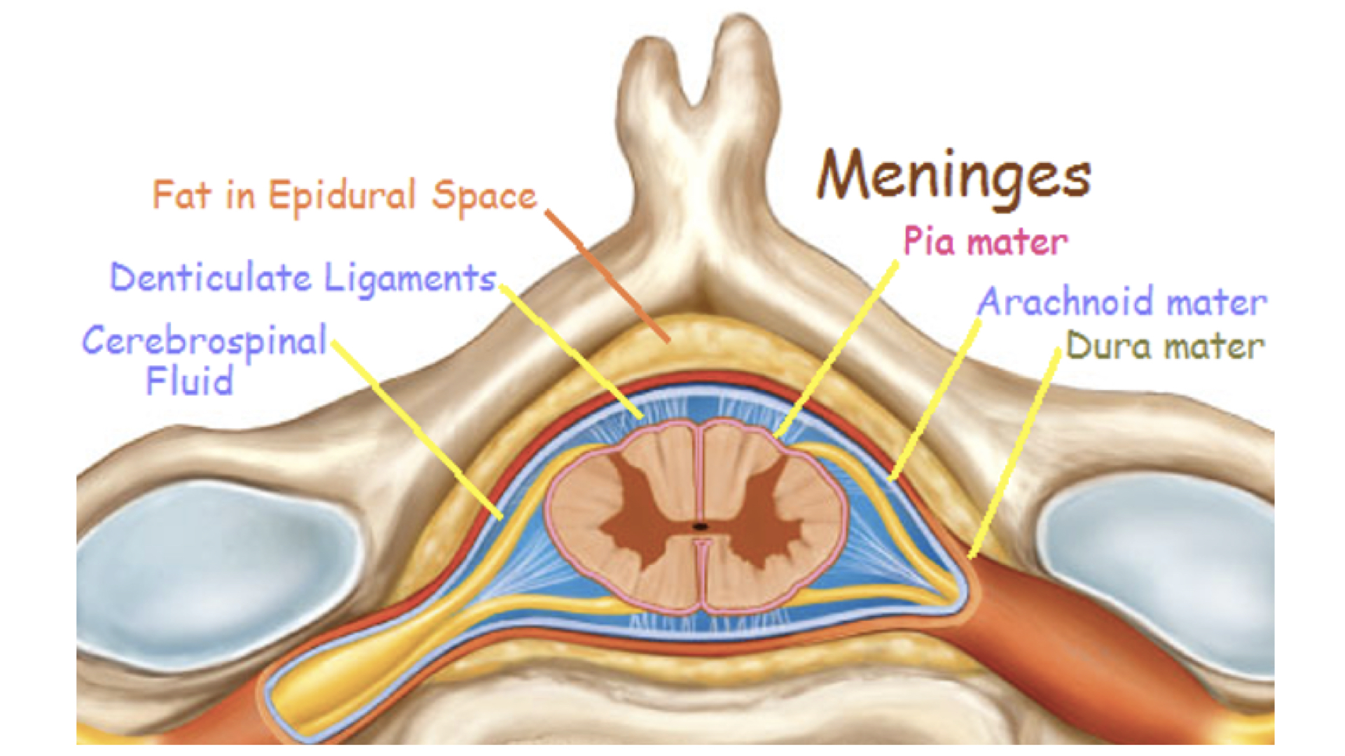
2. Covered with meninges - protective layers, prevent movement/shock
a. Dura Mater- “tough mother” dense connective tissue, covers spinal nerves to the intervertebral foramen, continuous with periosteum of the bone, creates on functional unit
b. Arachnoid – spider web, delicate connective tissue
c. Pia Mater – “delicate mother” – adheres to the surface of the brain and the spinal cord, can’t be removed. Contains many blood vessels for nourishment
d. Spaces
1. epidural – between the dura mater & vertebral column, contains blood vessels, adipose & loose connective tissue (provides padding around cord)
a. Epidural Block – anesthetic injected into the epidural space, affects only spinal nerves in immediate area, relieves pain
2. subarachnoid – space between the arachnoid and pia mater, filled with cerebrospinal fluid (CSF).
b. Spinal tap is a diagnosistic tool; fluid taken between L3-L4 as no cord there.
3. Cerebrospinal fluid –produced by the ependymal cells, in the choroids plexus located in the ventricles of the brain
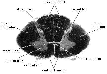
4. Composed of gray & white matter – histologically H shaped grey matter surrounded by white matter
a. Gray matter - unmyelinated tissue, mainly cell bodies, soma, nissl bodies
b. White matter - myelinated tissue, mainly axons
1. Produced by Schwann cells in PNS; oligodendrocytes in CNS
5. Denticulate Ligaments- help to hold the spinal cord in place, interweave with the periosteum of the bones
1. Central canal - contains cerebrospinal fluid – internal canal
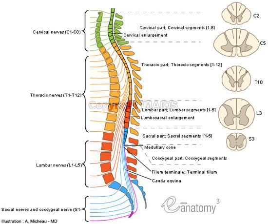
2. Conus Medullaris - tapered end at L1, L2 level – lumbar enlargement, length of cord is determined by age 4, it does not continue to grow
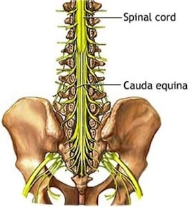
3. Cauda equina - fibrous strands extending from conus medullaris to coccyx ("horses tail”)--long ventral and dorsal roots.
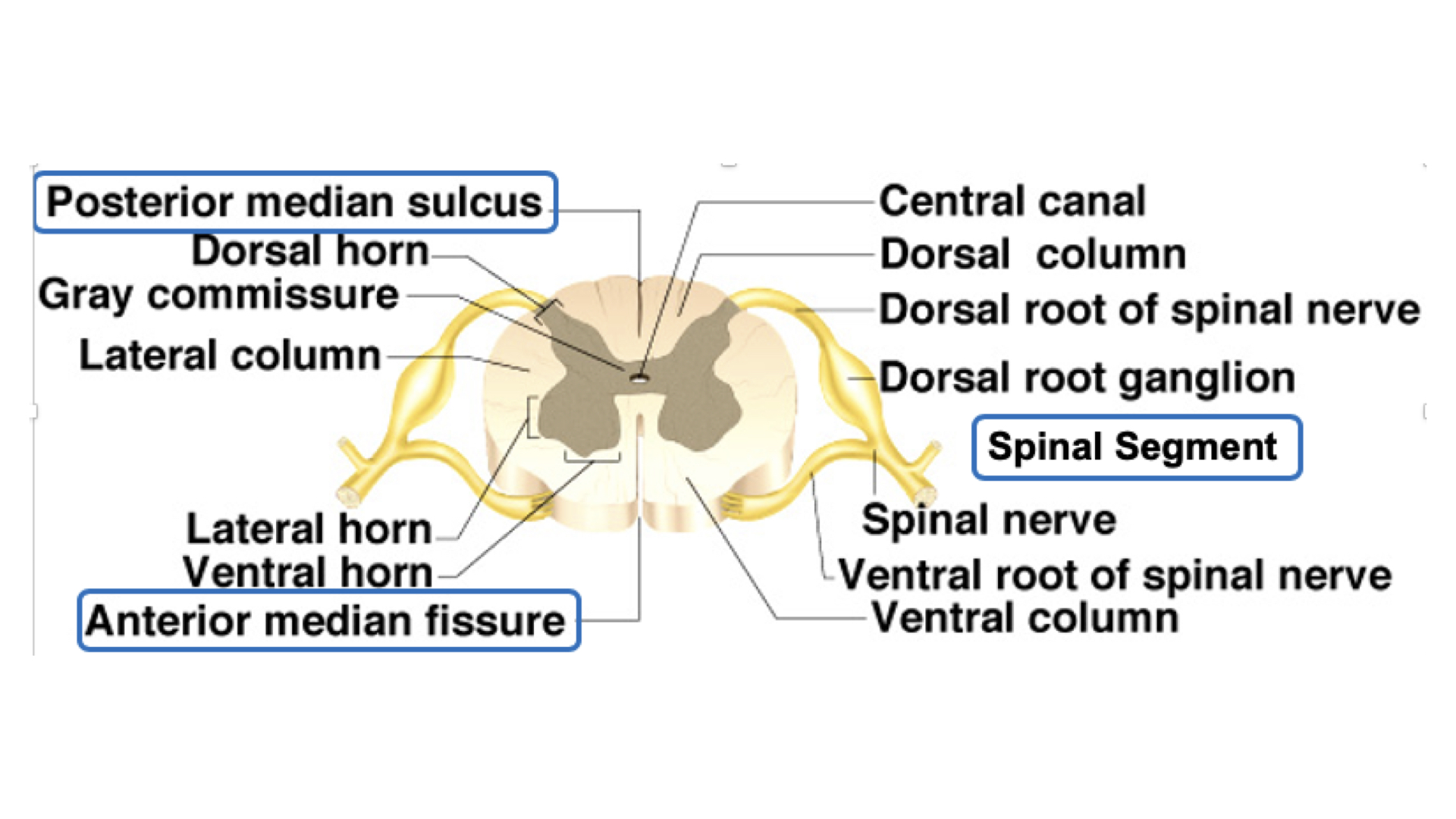
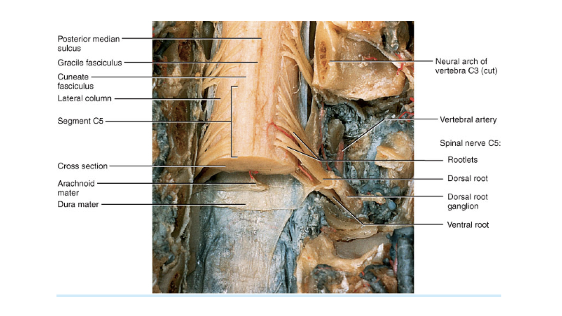
4. Anterior median fissure – deep groove along the ventral surface
5. posterior median sulcus – shallow groove on the dorsal surface
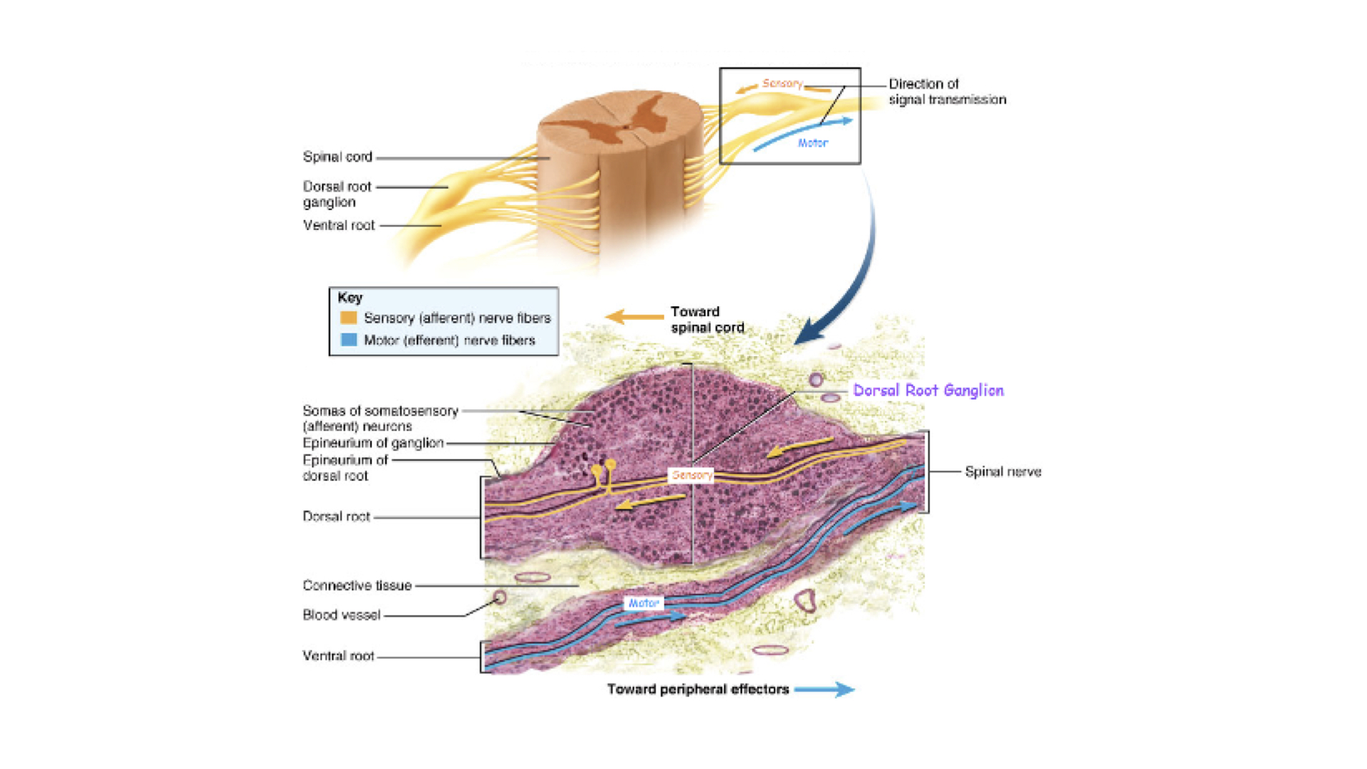
6. dorsal roots – sensory, afferent fibers, dorsal root ganglion, structurally unipolar, neurons from the periphery
7. ventral roots- motor, efferent fibers, dendrites and cell bodies in ventral horns, structurally - multipolar

1. Gray matter
a. Composed of neuron cell bodies,shaped like an H or butterfly
b. Six projections called horns:
1. Two dorsal gray horns
2. Two ventral gray horns
3. Two lateral gray horns - found in thoracic lumbar
c. Transverse bar called the gray commissure
d. Central canal in center of gray commissures
2. White matter
a. Contains two grooves that extend the length of the cord
1. Anterior median fissure
2. Posterior median sulcus
b. contains six fasciculi
1. Two posterior
2. Two lateral
3. Two anterior
c. Ascending and descending tracts are located in the fasciculi
1. share common origins, destinations, and functions
d. A column is a group of tracts
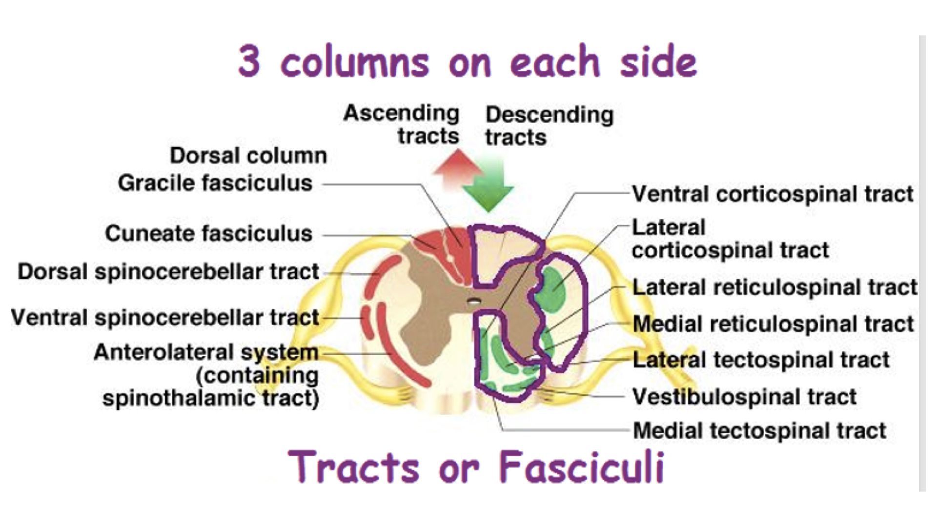
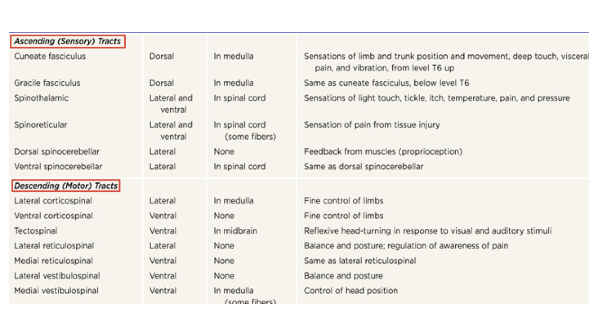
e. Pathways
1. Ascending pathways have three neurons from sensor to cerebral cortex. Note that all go through the thalamus which serves as a filter.
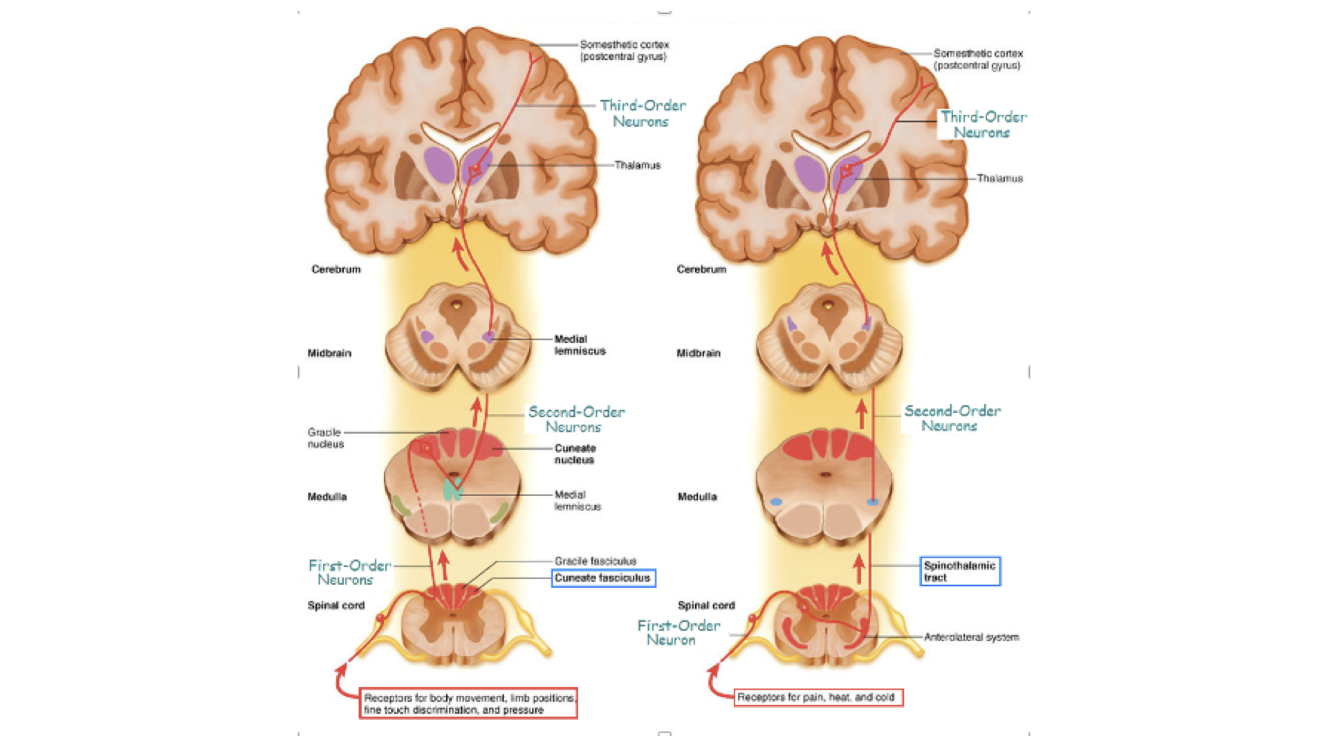 Ascending Pathways
2. Descending pathways have only two neurons between cerebral cortex and effector organ.
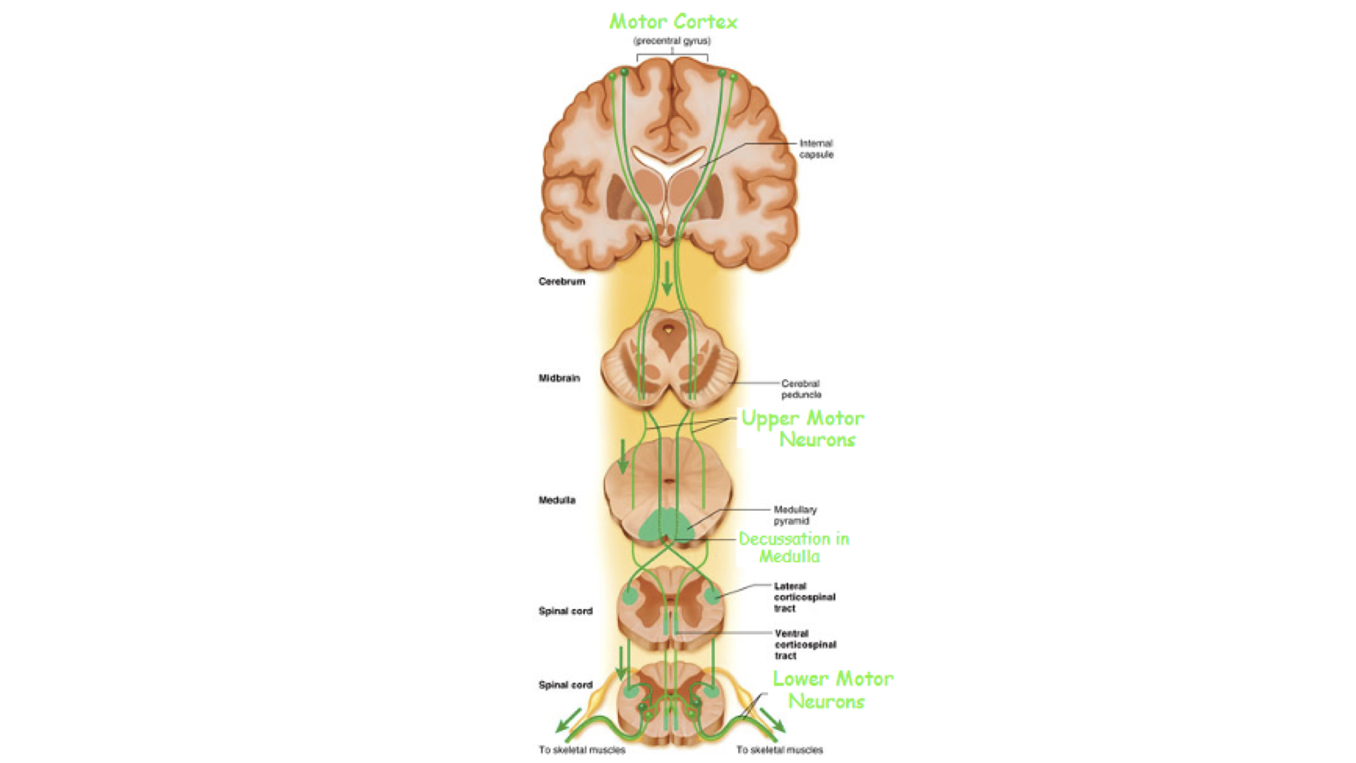
f. Decussation simply means the cross over between right and left. Remember that sensory data from right side of body and motor responses to the right side of the body involve the left cerebral hemisphere. Sensory and motor data from and to the left side of the body involve the right cerebral hemisphere.
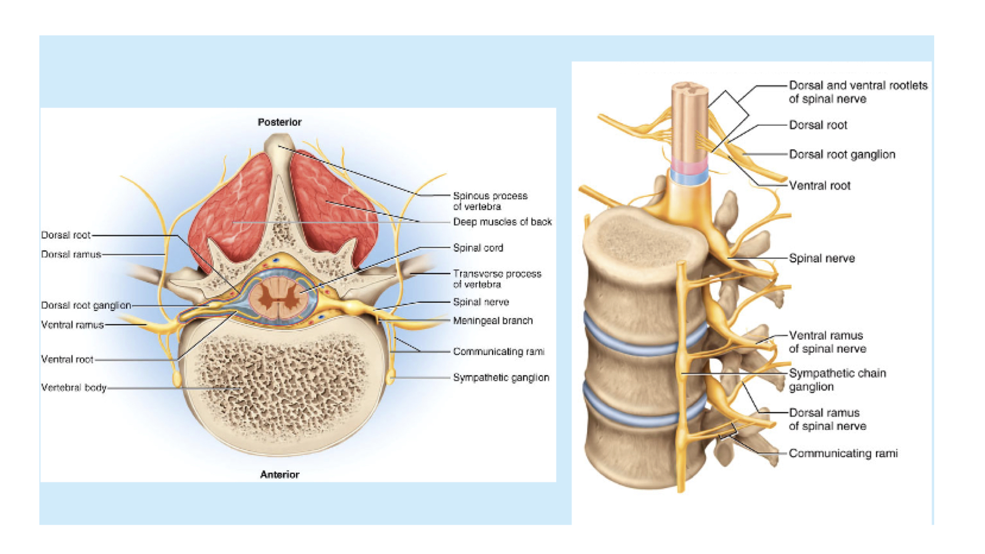
1. There are 31 pairs of spinals--one pair between each pair of vertebrae. these nerves are directed to specific areas of the body. Nerves are bundles of neurons just like muscles are buddies of muscle fibers. Spinals contain both sensory and motor neurons, Sensory neurons enter the spinal on the dorsal side--dorsal root. Most sensory neurons are unipolar and the cell bodies for each sensory neuron are found in a ganglion along the dorsal root.
Motor neurons have their cell bodies in the gray natter of the spinal cord and exit on the ventral side--ventral root. Just outside of the spinal cord, the spinal nerve is made up of both types of neurons.
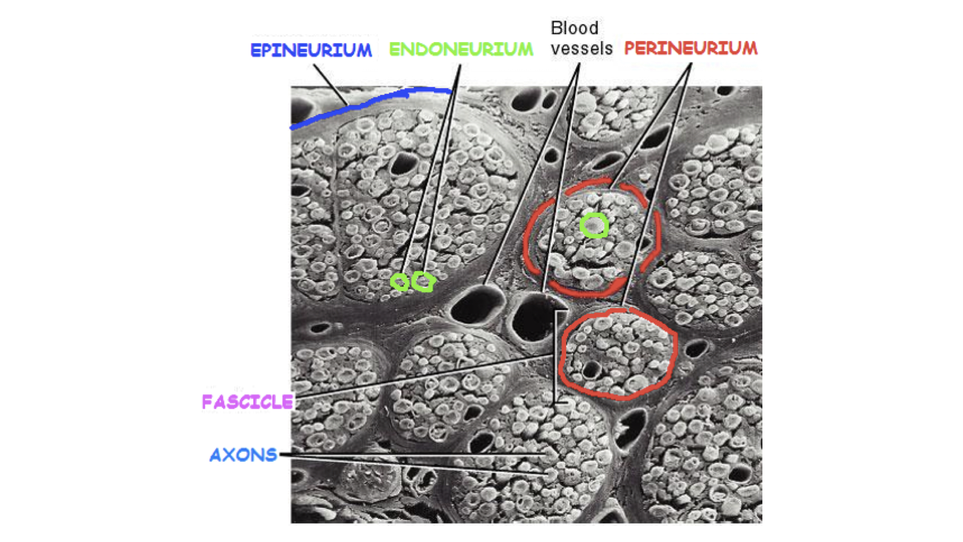
The nerve bundle has layers of connective tissue just like muscles did. The difference is that with nerves we use the prefix neuro-- instead of myo-.
Where the nerves enter and leave the spinal cord, separate not smaller bundles like a frayed rope. Here we call this rootlets.
 Mixed Spinal Nerve
2. After leaving the spinal cord, spinal nerves divide into dorsal, ventral, and communicating rami. The dorsal ventral simply go to those parts of the body at their respective level (recall dermatomes from the Integumentary System). The communicating rami integrate (communicate) with spinal nerves above and below the level.
3. Nerve plexuses are formed within each general area of the spinal cord except for the thoracic portion. In a plexus, nerves from different spinal levels integrate to form nerves that lead out into the body. You do not need to know all of those nerves. However, you do need to know the major nerve formed in each plexus.
a. Cervical plexus: The major nerve that is formed here is the phrenic nerve. The phrenic innervates the diaphragm and controls breathing.
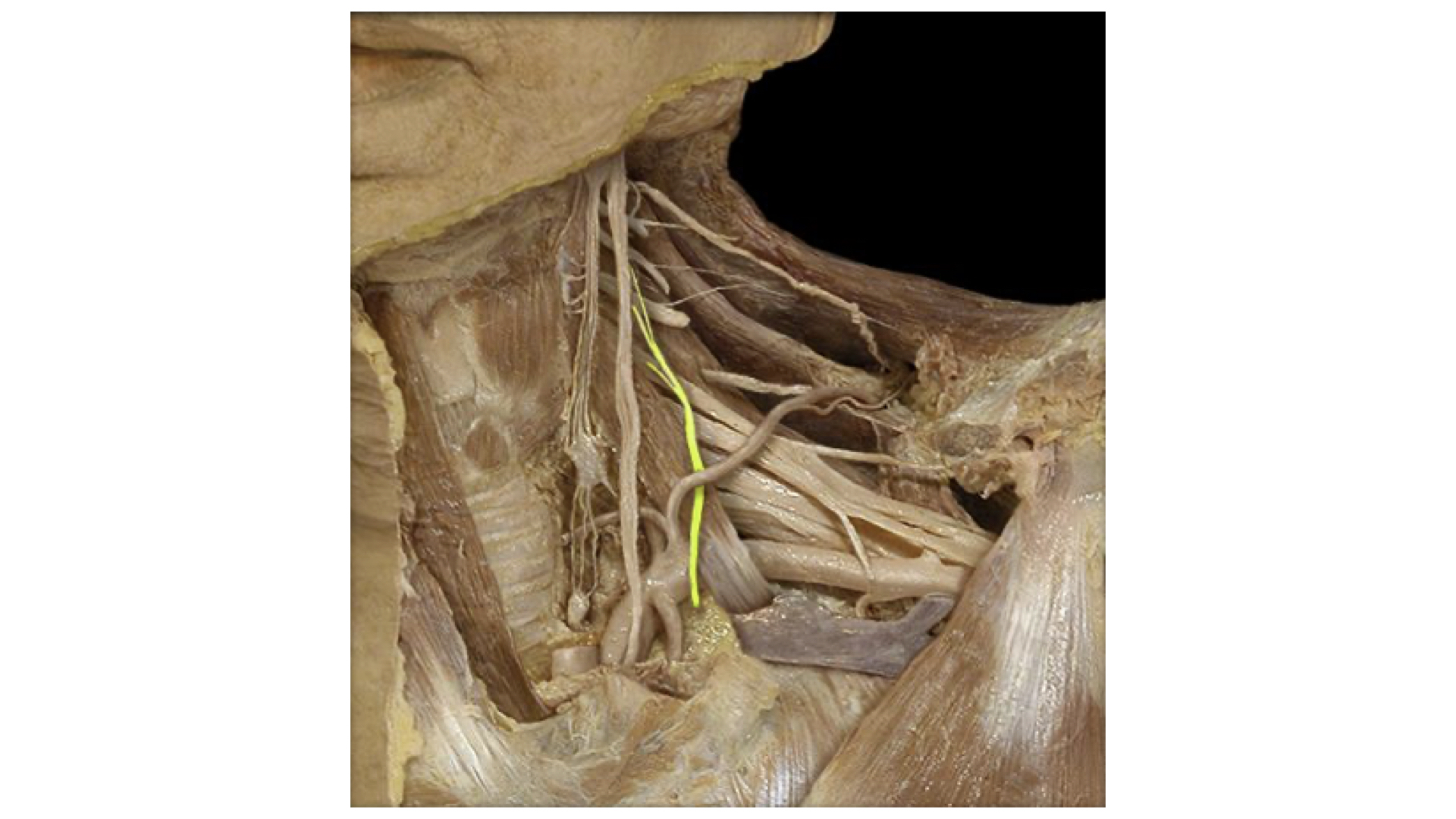 Cervical Plexus--Phrenic Nerve Highlighted
b. Brachial Plexus: The major nerves here are the radial, ulnar, and median nerves. The ulnar causes tingling down the forearm to the little finger when struck at the elbow--"crazy bone" response.
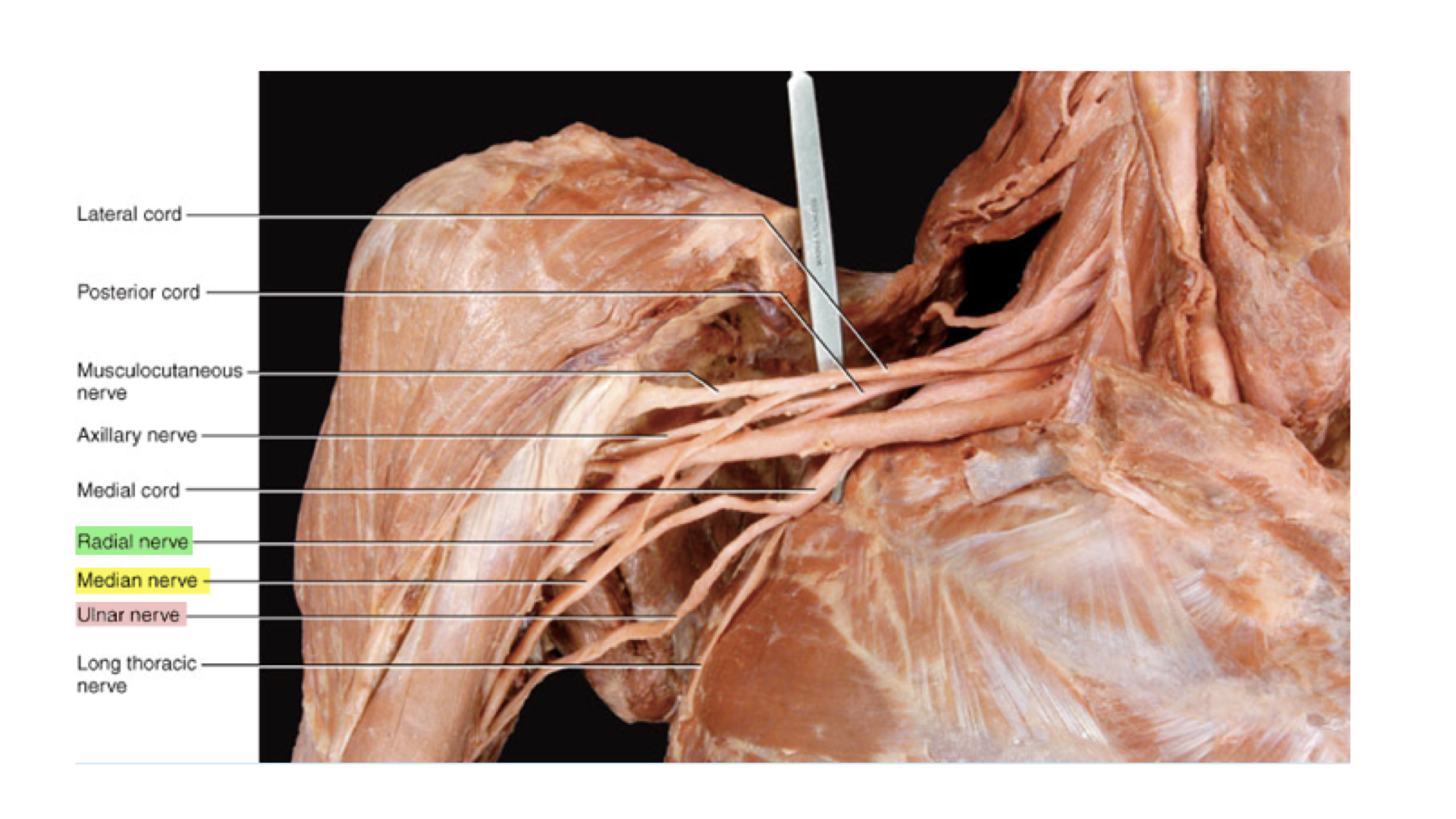 Brachial plexus with Radial, Ulnar, and Median Nerves Highlighted
c. No thoracic plexus,
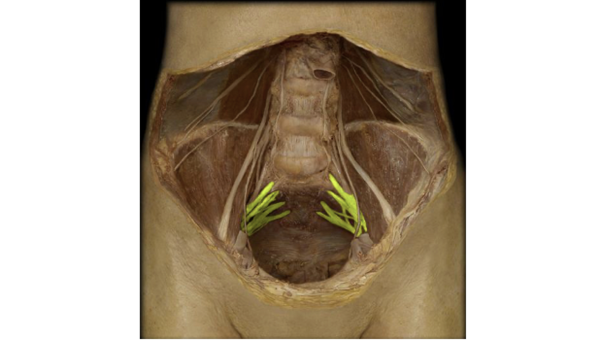
d. Lumbar Plexus. The major nerve from this plexus is the femoral.
 Lumbar Plexus with Femoral Nerve Highlighted.
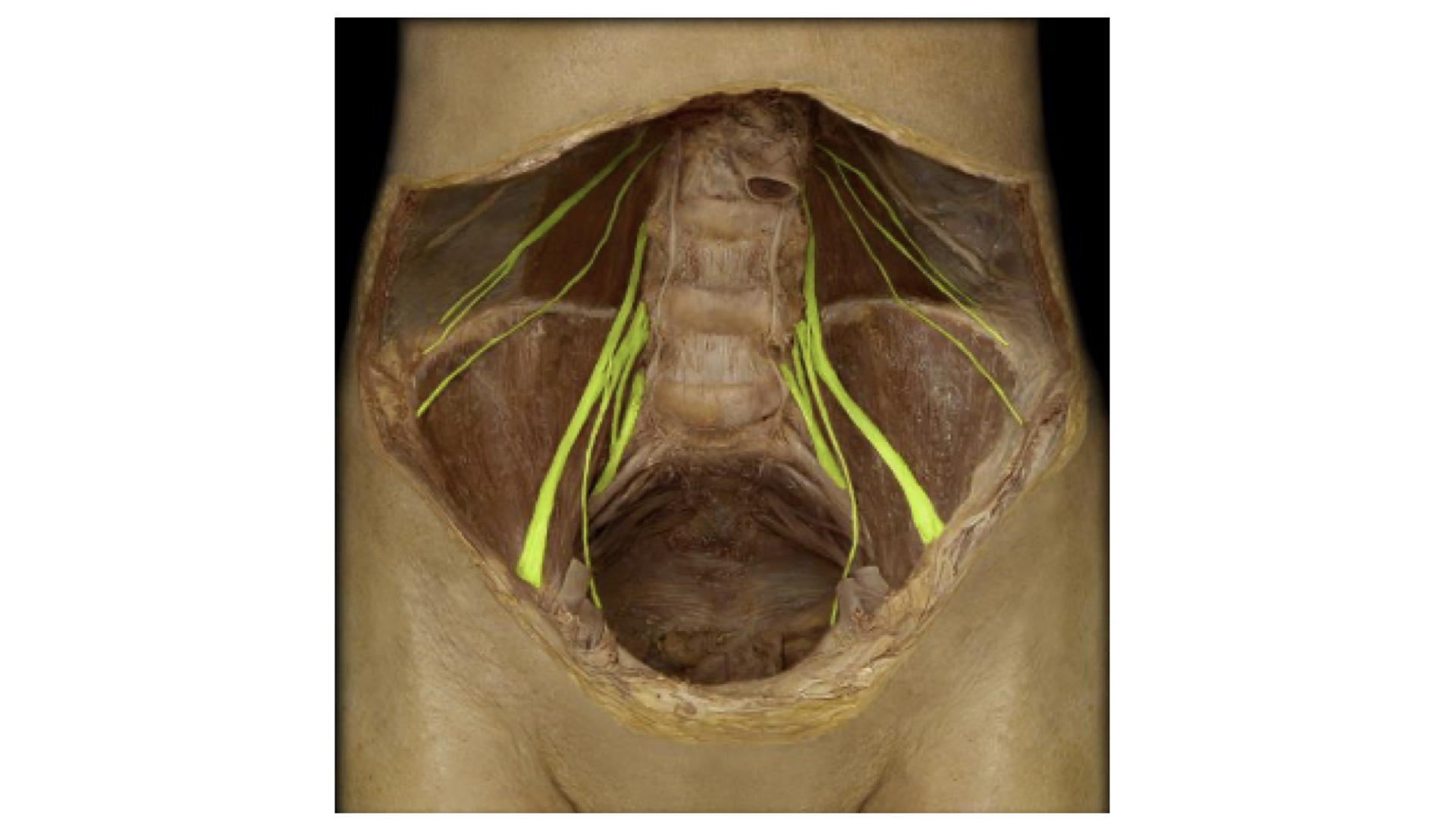
e. Sacral/Coccygeal Plexus: The primary nerve from this plexus is the sciatic nerve.
 Sacral/Coccygeal Plexus with Nerves forming the Sciatic Nerve Highlighted
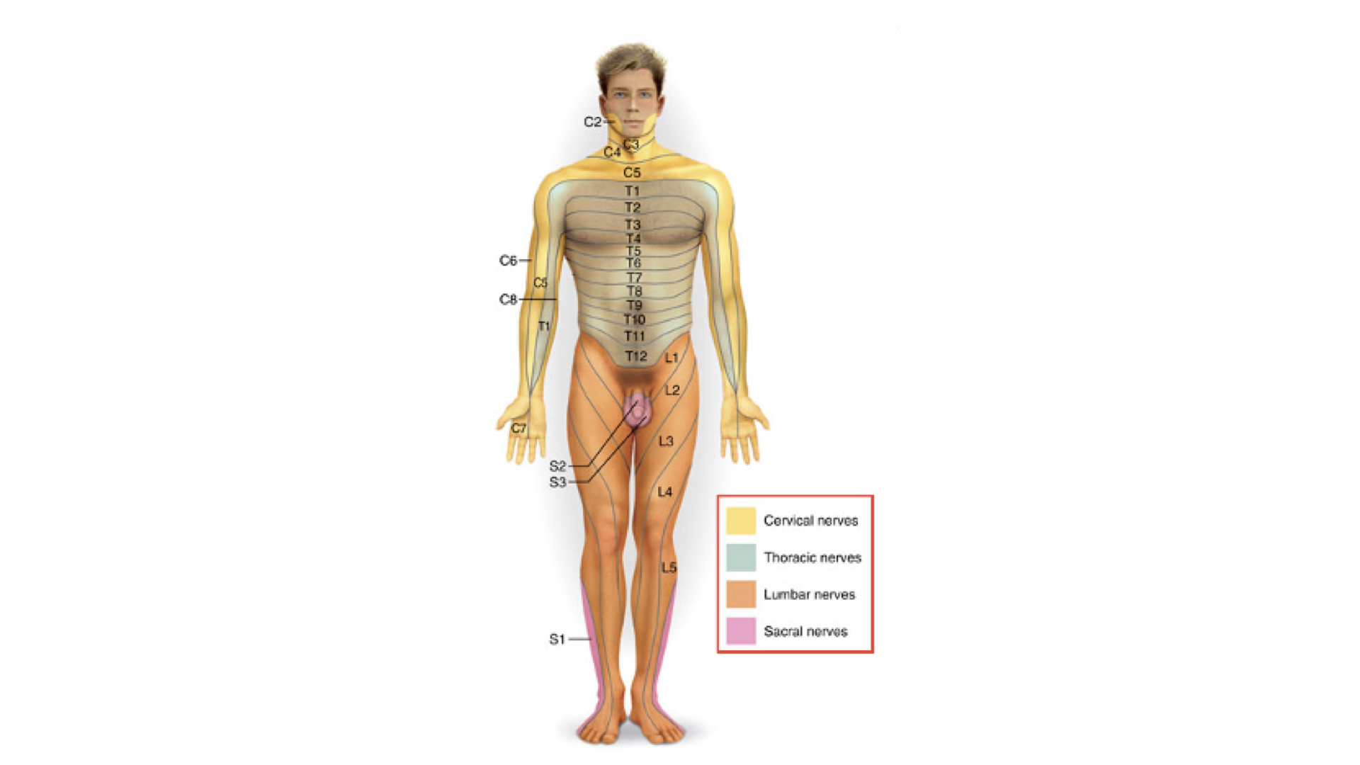
4. As mentioned above, spinal nerves innervate specific areas of the body. These areas are referred to as dermatomes. Below is a mapping of these dermatomes.
1. Automatic, unconscious, protective responses
2. Simplest type of nerve pathway
3. Sensory and motor impulses are not interpreted by the brain
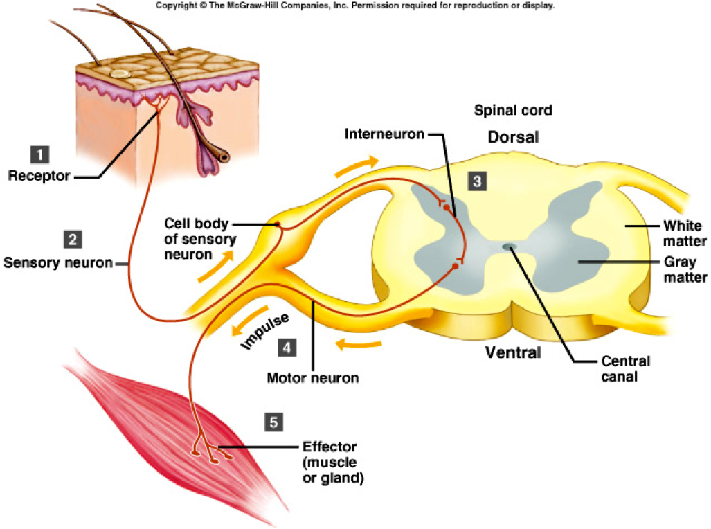
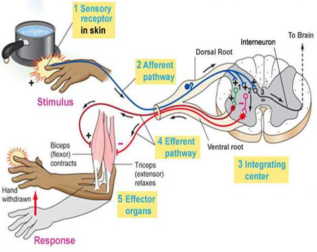
4. Steps involved with a reflex arc
a. Receptor picks up sensory impulse
b. Sensory impulse travels along spinal nerve, past dorsal root ganglion, through the dorsal root and enters the dorsal gray horn (same side of the body).
c. Sensory impulse is transferred to the ventral gray horn
1. Monosynaptic - directly from sensory to motor neuron-- most rapid
2. Polysynaptic - from sensory to interneuron to motor neuron--slower and more complicated response
d. Ventral gray horn sends motor impulses out of spinal cord, through the ventral root and along spinal nerves to muscles that will respond to the sensory stimulus
e. Effector responds.
f. Once initiated, a reflex cannot be stopped. Examples :
1. Sneezing
2. Coughing
3. Gag reflex
4. Patellar reflex
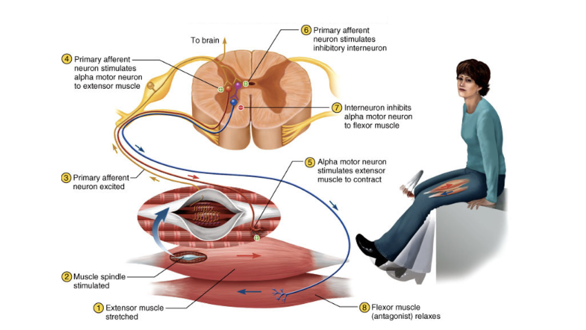
a. Reciprocal innervation
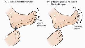
5. Babinski reflex / Plantar reflex
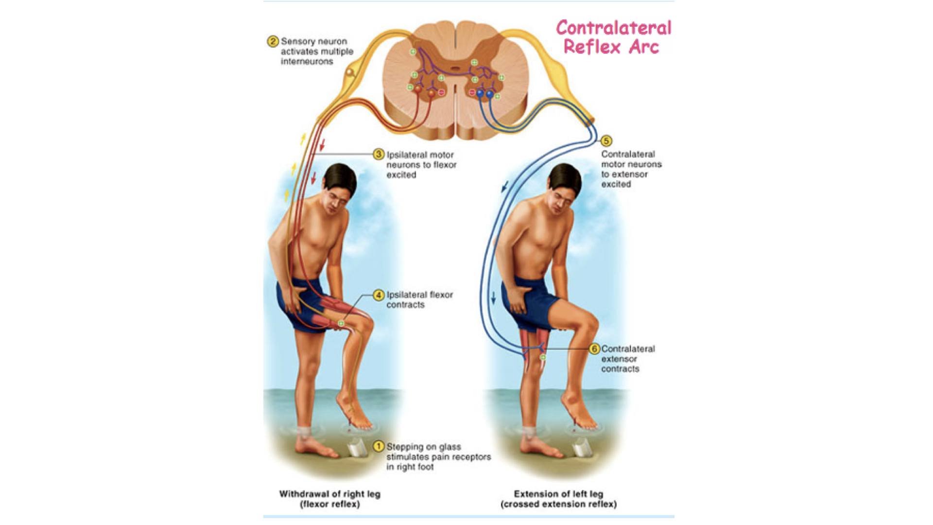
6. Flexor reflex-- withdrawl
7. Crossed extensor reflex
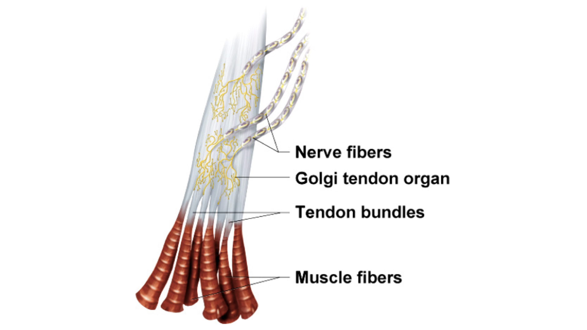
8. Goigi Tendon Reflex - When tendons become overstretched, reflex causes relaxation of the muscle involved.