Structure and Function of the Digestive System
Structure and Function of the Digestive System
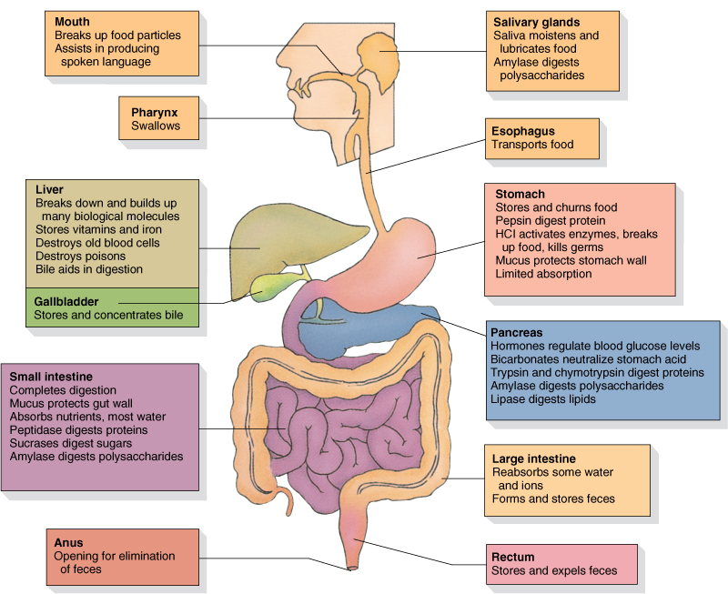
Digestive function
1. Ingestion - This is an active process that includes both mechanical and chemical processing. It is also a conscious choice
2. Mechanical processing -This includes chewing and mixing of materials in the stomach.
3 Digestion - This includes chemical breakdown of food into organic fragments
4. Secretion - This includes release of water, acids, enzymes, buffers and salts into the appropriate areas of the system--Mouth, Stomach, and Small Intestine.
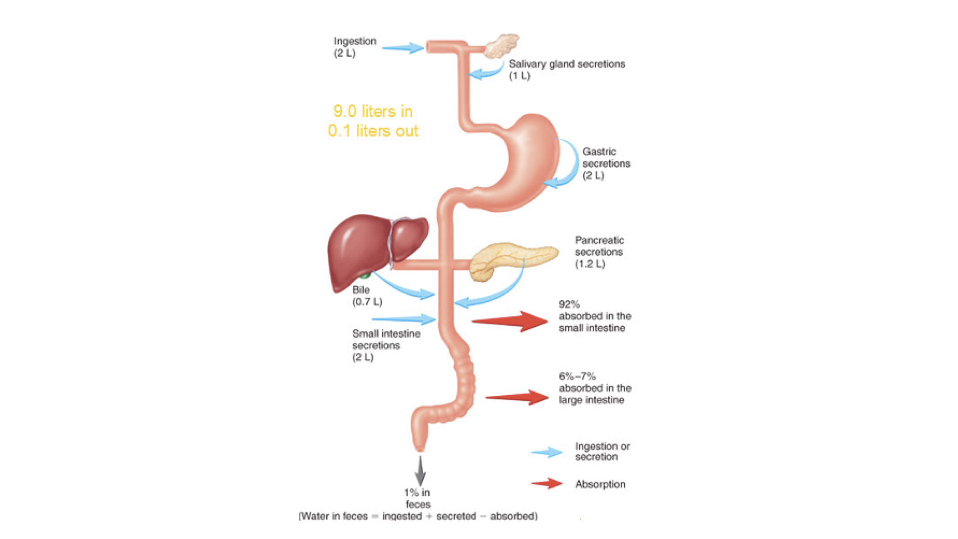
5. Absorption - This includes movement of organic substrates, electrolytes, vitamins and water across the epithelium into the interstitial fluid of the digestive tract. From there, they go into the blood or lymph to be transported.
6. Excretion - Elimination of waste products. This includes defecation which simply the elimination feces. Some materials absorbed are processed in the liver and filtered out in the kidneys. They are eliminated in the urine via urination/ micturition.
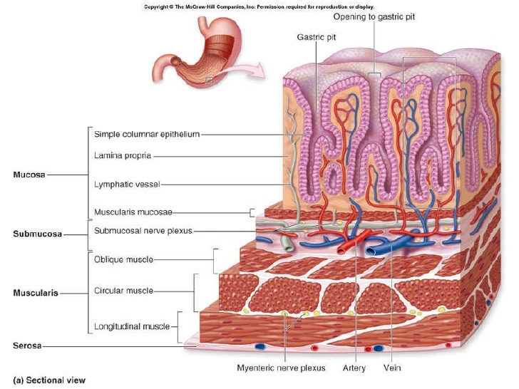
There are four layers of tissues in the digestive organs.
1. Mucosa - The inner lining is composed of epithelial tissue that contains glands that secretes mucous, enzymes and bicarbonate ions. The lamina propria is the underlying layer of loose connective tissue. This layer contains blood and lymph vessels.
2. Submucosa - This layer is a dense connective tissue which also contains nerve plexuses of the enteric nervous system along with arterioles and venules.
3. Muscularis - This layer is made of smooth muscle. The muscles may be in a circular pattern or longitudinal. Contraction facilitates mechanical processing and movement of materials along the tract. Some of these are pacesetter cells that can spontaneously trigger action potentials and contractions in the muscle. Those movements that propel materials through the digestive tract are called peristalsis. Segmentation occurs in most areas of small intestine , some areas of the large intestine
which facilitate with churning of contents to mix with secretions
4. Serosa - This layer makes up the serous membrane. It has both a visceral layer covers the muscularis externa and the parietal layer which is attached to the abdominal wall.
a. mesenteries - double sheets of loose connective tissue
Functions
- Mechanical processing
- Lubrication
- Analysis
- Limited digestion of carbohydrates
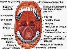
Anatomy
- Hard palate - palatine process of maxillary bone
- Soft palate - posterior to hard palate uvula-
- Tounge - skeletal muscle
- Salivary glands - 3 pair - each produces a slightly different type of saliva
a. salivary amylase - parotid salivary glands breakdown complex carbohydrates into maltose
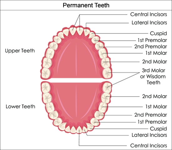
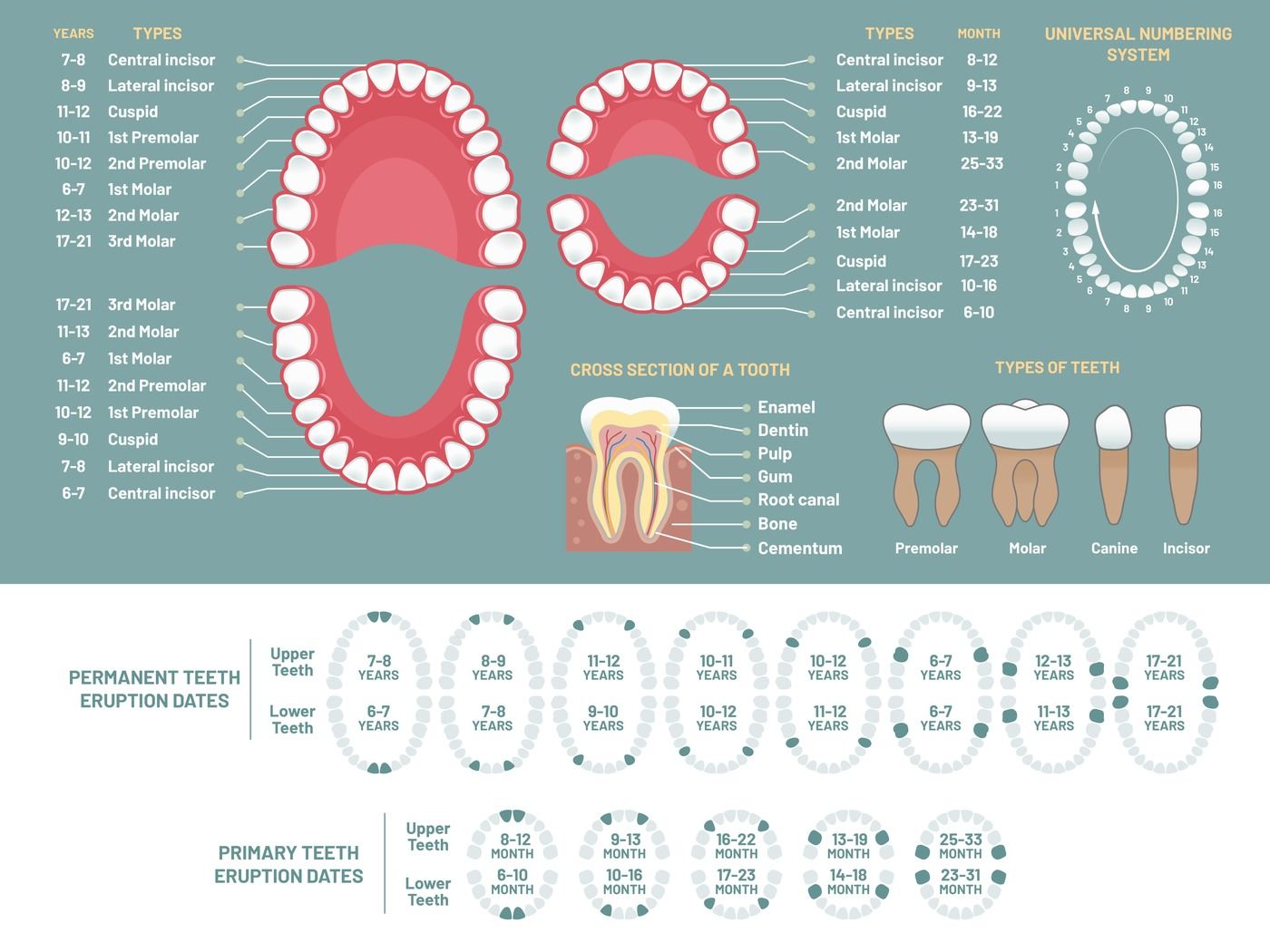
Teeth
- Mastication--chewing
a. Crown - calcium phosphate
1. Enamel - hardest biological substance manufactured in the body-- Fluoride
2. Dentin - does not contain living cells
3. Pulp - cytoplasmic processes extend into the dentin
b. Neck - boundary between the crown and the root
1. Gingiva - gums
2. Gingivitis
c. Root - gomphosis joint joins the dentin of the root to the avelolar bone
1. Root canal
2. Cementum - resembles bone but does not remodel like bone
d. Deciduous teeth - baby teeth, milk teeth, primary teeth
Secondary dentition - permanent teeth
1. Incisors
2. Cuspids
3. Bicuspids
4. Molars
e. Saliva contains salivary amylase which begins digestion of carbohydrates.
Pharynx - common passageway for food , liquids, and air
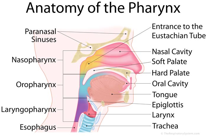
Esophagus - muscular tube to carry food and liquids to the stomach.
1. Swallowing - deglutition
a. bolus - oval shaped ball of food
b. Hiatal hernia - abdominal organs are pushed through the esophageal hiatus
2. Solids and liquids pushed through esophagus by peristalsis.
The stomach
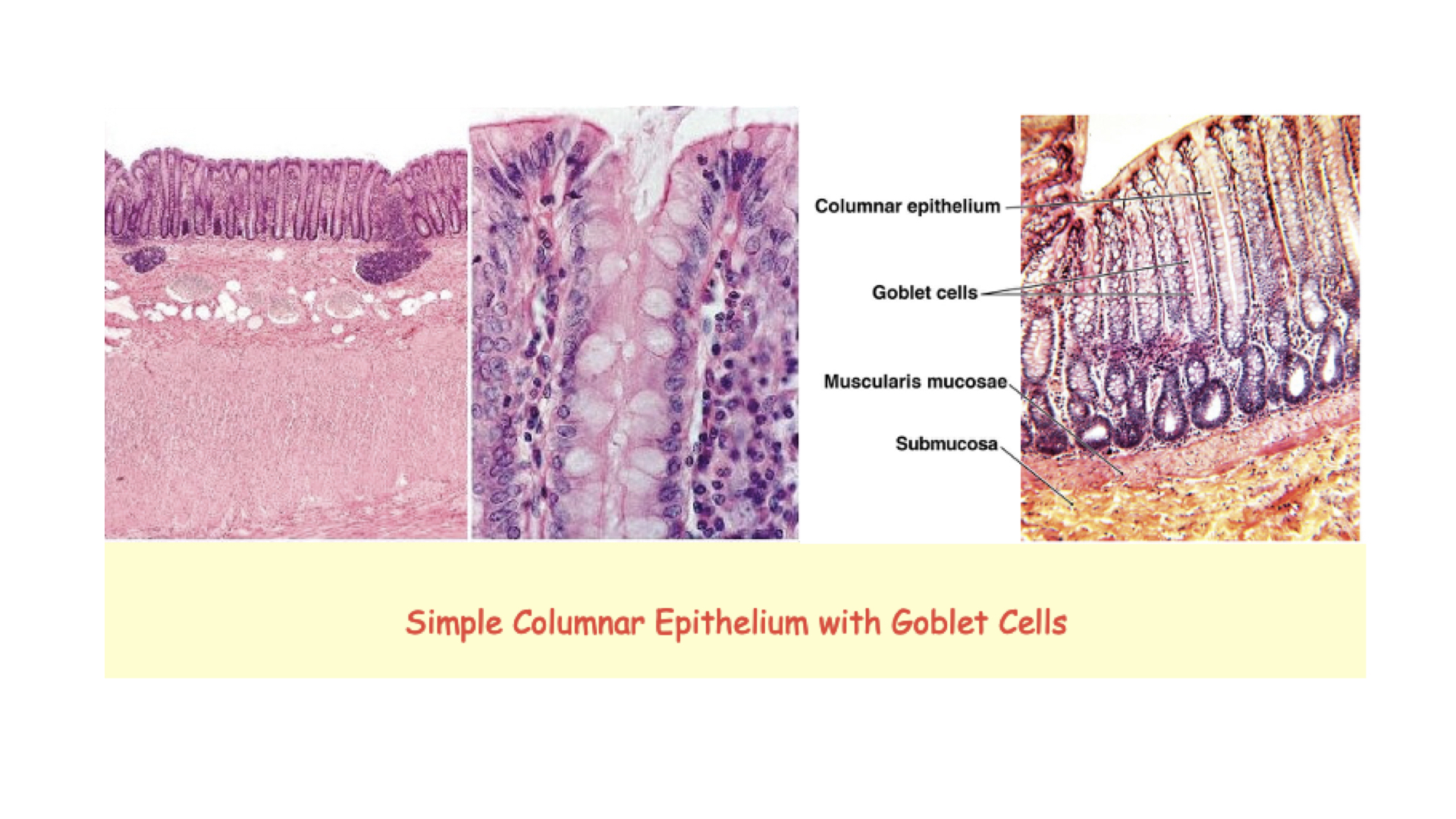
1. Lined with simple columnar epithelium
Major Functions
1. Bulk storage
2. Mechanical breakdown
3. Use of acids and enzymes to breakdown chemical bonds
4. Production of the intrinsic factor - glycoprotein necessary for the absorption of vitamin B12
5. Chyme - ingested materials mixed with gastric juices
Anatomy

Four regions of the Stomach
1. cardia
2. fundus
3. body
4. pylorus
a. pyloric sphincter
b. Rugae - ridges and folds
Muscles of the stomach---circular, longitudinal, oblique
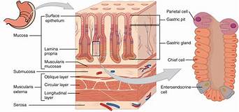
Gastric wall
1. Alkaline mucous layer - produced by the epithelium to protect against the acids and enzymes2. 2. Gastric pit - shallow depression
3. Gastric gland - located in the fundus and body of the stomach
a. Parietal cells
1. intrinsic factor - glycoprotein that facilitates the absorption of B12
2. Hydrochloric acid (HCL)
a. kills most microorganisms
b. denatures proteins
c. activates pepsinogen to pepsin
d. breaks down plant cell walls and connective tissue in meat
b. Chief cells secrete pepsinogen
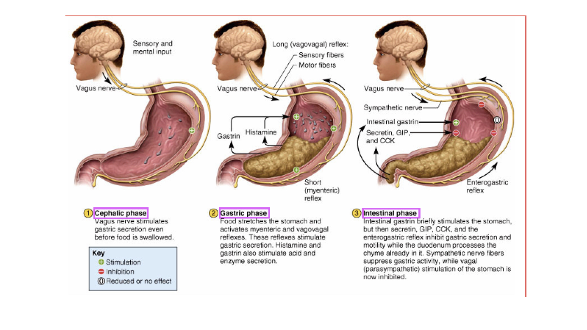
Phases of Gastric Activity
1. cephalic phase: prepares stomach to receive food.
a. Controlled by autonomic nervous system
1. Parasympathetic through vagus nerve increases activity
2. Sympathetic nervous system through coeliac ganglion decreases activity
2. gastric phase - activated by
a. arrival of food
b. increase pH
c. distention of stomach
d. gastrin - hormone - stimulates contractions of smooth muscles
3. intestinal phase - when chyme enters small intestine, controls the rate of gastric emptying
a. intestinal hormones
1. secretin is released by pancreas when chyme enters the duodenum
2. cholecystokinin (CCK)
3. gastric inhibitory peptide (GIP)
Digestive Activity in Stomach
1. Pepsin begins digestion of proteins--including salivary amylase.
2. Hydrochloric acid breaks cells apart and kills most bacteria.
3. Absorption of aspirin and some lipid-soluble drugs.
Ulcers
1. Normally the stomach wall is protected by mucous and continual cell replacement. When this protective layer of mucous is eroded, open sore can develop.--ulcer.
Anatomy
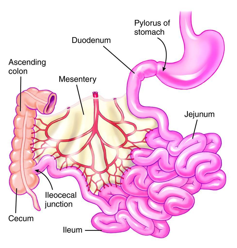
- Duodenum - closest to the stomach
- Jejunum - most of chemical digestion and nutrient absorption occurs here
- Ileum - ileocecal valve - controls flow into large intestine
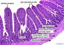
Intestinal wall anatomy
1. picae - transverse folds
2. villi - finger like projections
3. microvilli - cover each villus
a. lacteal - terminal lymph - located in each villus
1. chylomicrons - protein-lipid packages too large to enter blood capillaries travel through lymph
Intestinal Secretions
1. Moistens the chyme and buffers the acids from the stomach
a. Wall of small intestine is not protected from acidity
2. See sections on liver and pancreas for specifics of enzyme secretions
 Digestion of Carbohydrates
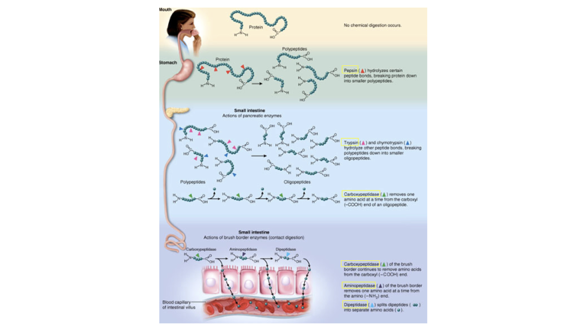 Digestion of Proteins
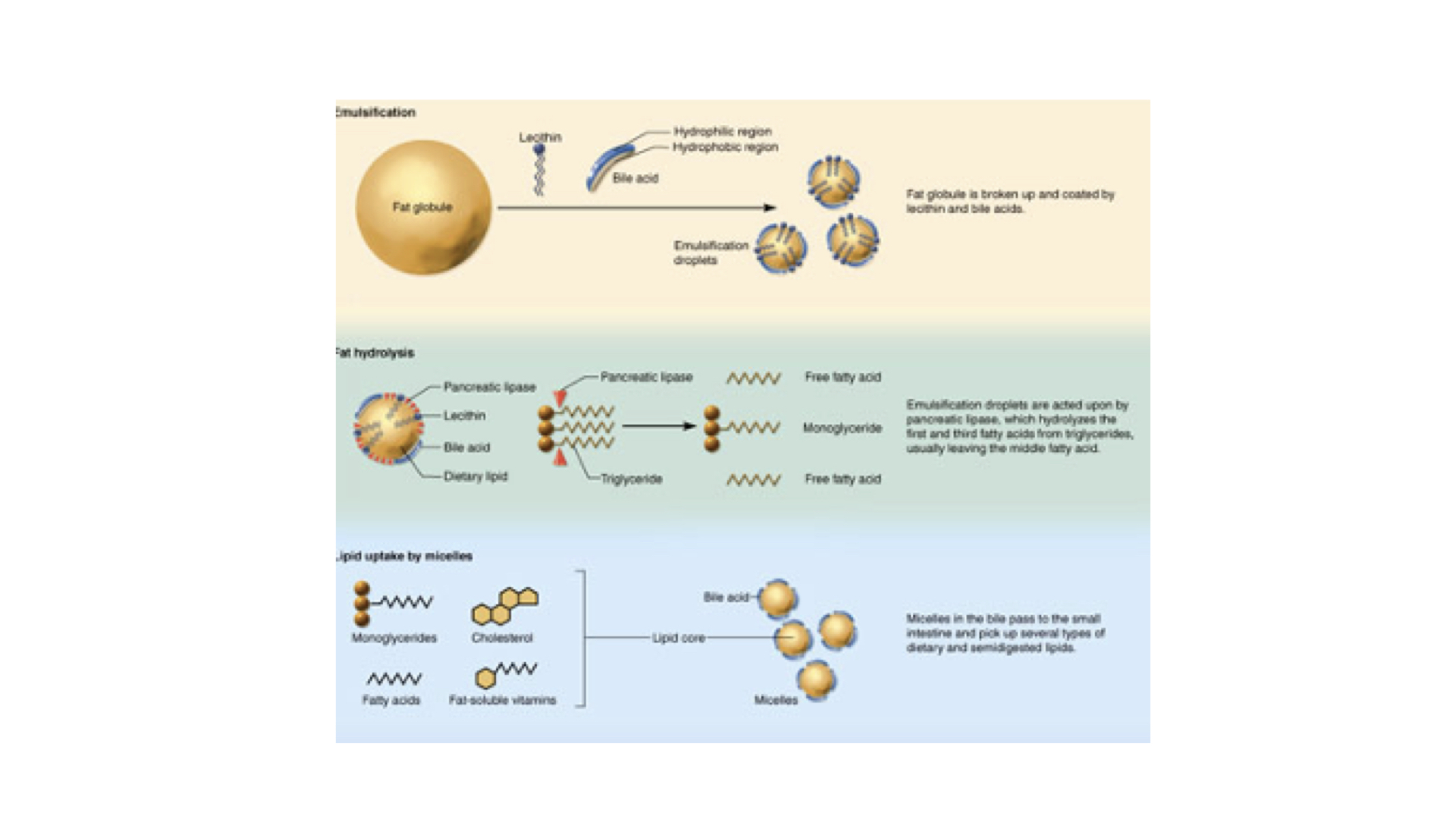 Digestion of lipids
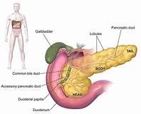
The Pancreas- endocrine/exocrine organ
1. Pancreatic islets
a. alpha cells - Produce glucagon
b. beta cells - Produce insulin
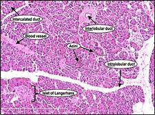 Pancreatic Histology
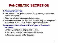
2. Acinar cells - secrete pancreatic proenzymes
a. Carbohydrase - alpha-amylase
b. Lipases
c. Proteases
d. Peptidases
Functions
1. Metabolic regulation
a. Extract absorbed nutrients or toxins
b. Monitor and adjust circulating levels of organic nutrients
c. Fat soluble vitamins A,D,E,K are absorbed and stored
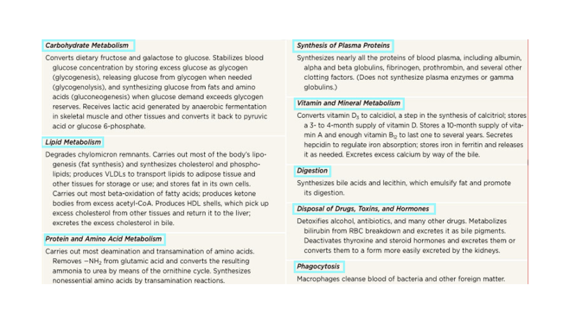
2. Hematological regulation - largest blood reservoir in the body
a. Synthesize plasma proteins
3. Bile production - synthesized from cholesterol
a. Normal digestion and absorption of fats
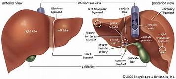
Anatomy - Four Lobes
1. Left
2. Right
3. Caudate
4. Quadrate
Liver lobule - basic functional unit
1. Hepatocytes
2. Sinusoids - specialized highly permeable capillaries empty into a central vein.
a. Kupffer cells are located here
3. Common hepatic duct - bile leaves the liver and goes to
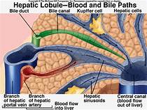
Double Blood Supply
1. Hepatic artery - oxygenated blood
2. Hepatic portal vein - deoxygenated blood carries newly absorbed nutrients to liver
Anatomy
Function
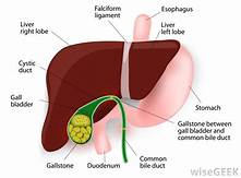
1. storage area for bile
2. modification of bile
Bile Function
1. emulsification - bile salts (assortment of lipids) break apart, large droplets of lipids, then coat them to make it easier for enzymes to attack
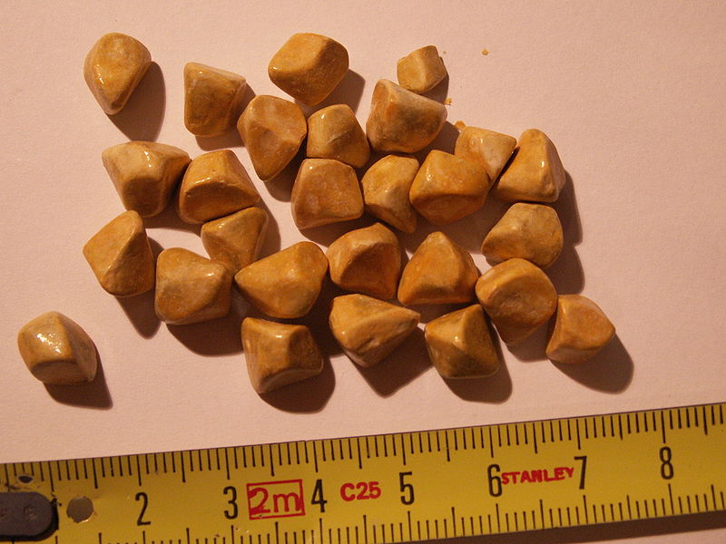
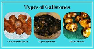
2. Gallstones - stored bile becomes concentrated
Functions
1. Reabsorption of water
2. Compaction of feces
3. Absorption of vitamins
4. Storing of fecal material
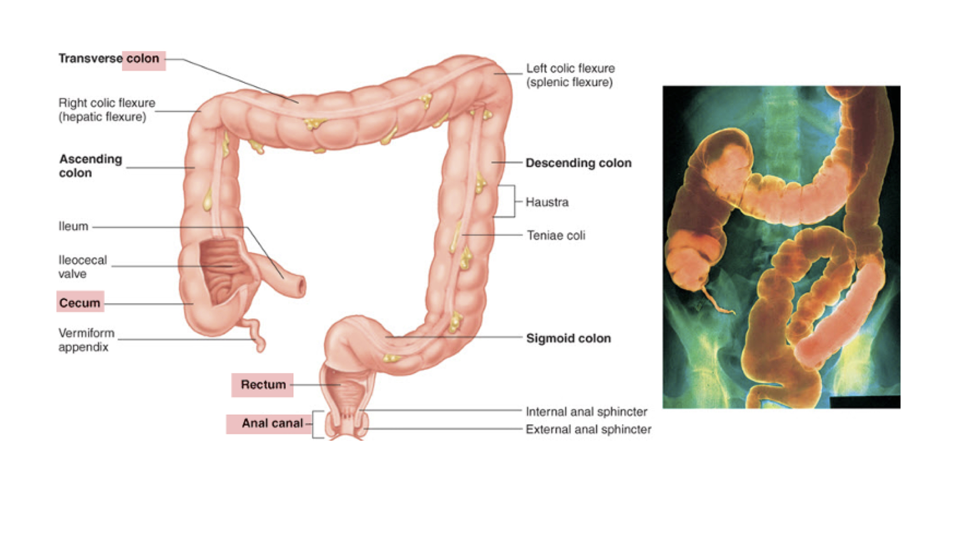
Anatomy
1. Colon
a. Ascending
b. Transverse
c. Descending
d. Sigmoid
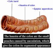
e. Haustra - pouches that contract independently to move material to next pouch
2. Cecum
a. Ileocecal valve - Regulates movement of material from ileum into ascending colon
b. vermiform appendix - Blind pouch at end of cecum
1. Function unclear
3. Rectum
4. Anus
Physiology of the Large Intestine
1. reabsorption of water-important to retain water added to food as digestive fluids
2. Bacteria - synthesize vitamin K, biotin, and vitamin B5
3. decompose bilirubin (breakdown product of hemoglobin) into simpler products
4. Toxins - proteins are broken down into amino acids – recycled or excreted, by-product may be hydrogen sulfide gas
5. Carbohydrates are fermented - release H2, CO2 and methane gas
Common Disorders
1. Diarrhea
2. Constipation
3. Colon cancer - Colonoscopy done depending on age and family history