How the Respiratory System Works
How the Respiratory System Works
We normally think of respiration as the movement of air into and out of the lungs. This action should not to be confused with cellular level respiration that occurs inside of cells to produce ATP, although both are needed along with proper functioning of the cardiovascular system for you to stay alive. Any disruption of these results in a loss of homeostasis and death of the organism. The Respiratory System is involved in more than gas exchange. Functions include:
- Pulmonary ventilation - oxygen/carbon dioxide exchange.
- Vocalization in general and speech in particular.
- Supports olfaction by bringing air to the olfactory receptors.
- Control of pH by eliminating carbon dioxide.
- Assist with blood pressure regulation through angiotensin II
- Changes in pressure in the thoracic cavity assist venous blood return and movement of lymph.
Lumen Learning has several pages devoted to this system
Another good link to the human respiratory system is at the Online Biology Book
Typically the respiratory system is divided into an upper and lower system. The upper system is often referred to as the conducting zone because it is the path by which air gets into and out of the lungs. The lower half is referred to as the Exchange Zone because oxygen and carbon dioxide are exchanged here.
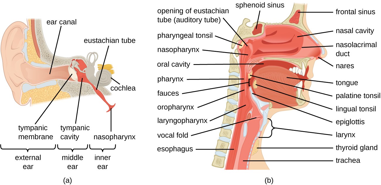 Upper Respiratory System
External nose - flexible cartilage
Nasal cavity
a. From nares to choanae
b. Vestibule: just inside nares
c. Hard palate: floor of nasal cavity
d. Nasal septum: partition dividing cavity. Anterior cartilage; posterior vomer and perpendicular plate of ethmoid
e. Choanae: bony ridges on lateral walls with meatuses between. Openings to paranasal sinuses and to nasolacrimal duct
Functions - 1. incoming air is warmed, moistened and filtered – nasal conchae - superior middle inferior
a. ciliated epithelium
b. mucous glands
2. olfactory stimuli are received
3. large hollow resonating chambers are provided for speech sounds – sinuses- frontal, maxillary, sphenoid, ethmoid
Pharynx –(throat) shared by the respiratory and the digestive systems
1. nasopharynx – to posterior edge of soft palate
a. Openings of Eustachian (auditory) tubes
b. Uvula is posterior extension of the soft palate.
2. oropharynx – soft palate to hyoid bone
3. laryngopharynx – hyoid bone – esophagus
Functions - 1. passageway for air and food
2. provide a resonating chamber for speech sounds
3. Tonsils – lymphoid organs –pharyngeal (adenoids when inflamed) and two lateral palatine tonsils
Larynx - voice box
Functions 1. connects pharynx with the trachea
2. contains 9 cartilage's - epiglottis - prevents entry of food or liquid into the respiratory tract – Box shapedThyroid cartilage →ridge →Adam’s Apple
3. sound production - vocal cords vibrate when air passes through the glottis – vocal cords = fibroelastic bands – length changes at puberty, larynx in male enlarges more = deeper voice
Trachea - windpipe
Functions 1. provides a tough flexible tube for passage of air
a. composed of 16-20 "C" shaped cartilage
b. opened part allows for expansion
c. lined with pseudostratified ciliated epithelium
Carina - cartilage at bifurcation. Membrane of carina especially sensitive to irritation and inhaled objects initiate the cough reflex
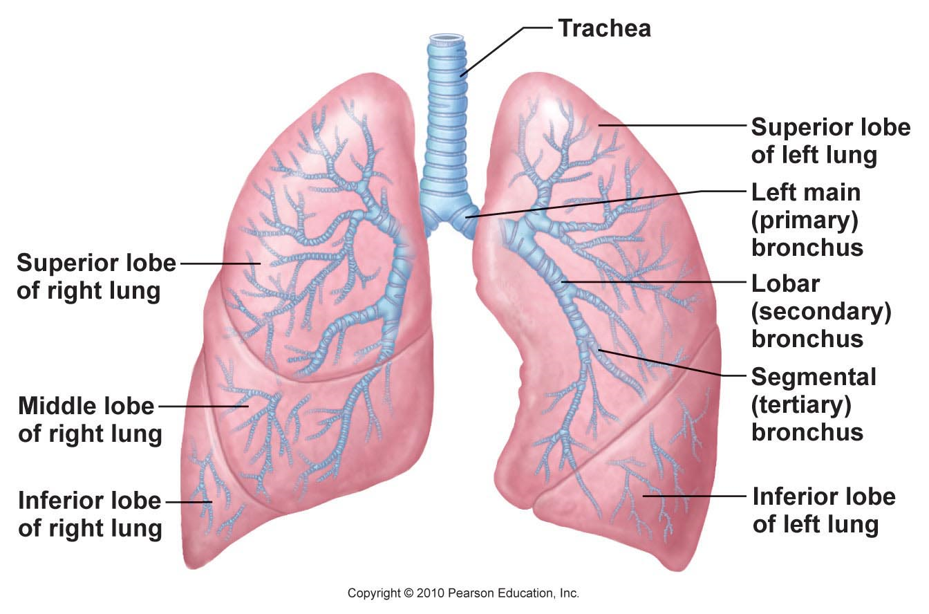
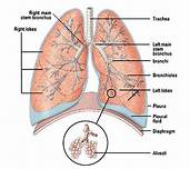
Lungs - gross anatomy
Pleural Cavity – each lung has it’s own cavity separated by mediastinum
1. Parietal pleura – inner surface of thoracic cavity, diaphragm, & mediastinum
2. Visceral pleura – outer surfaces of lungs & between the lobes in the fissures
Paired organs - - lobes separated by deep fissures
Apex - superior point of lung
Base - rests on the diaphragm
Hilus - on medial surface where bronchi and blood vessels enter the lung. All the structures in hilus called root of the lung.
Right lung - 3 lobes – superior, middle, inferior
Left lung - 2 lobes, - superior, inferior, cardiac notch
Bronchi - bronchial tree
Structure 1. Right and left primary bronchi
2. secondary bronchi - enter each lobe of that lung
3. tertiary bronchi - 9-10 divisions
4. bronchioles - can vary in diameter – smooth muscle
5. terminal bronchioles - provide passageway to the respiratory bronchioles
6. alveolar ductsalveolar sacsalveoli - the exchange surface of the lungs
Surfactant - oily secreation that coats the alveolar epithelium
1. reduces surface tension within the alveolus prevents the collapse of aveolar walls
Respiratory Membrane - gas exchange area
Structure 1. surfactant --> alveolar epithelium ( simple squamous ) -->fused basement membrane -->endothelium of the capillary ( simple squamous
Function - place where large quantities of air and large quantities of blood come in close contact for rapid exchange of gases to occur -diffusion – of gases – area of high concentration to area of low concentration