Blood Vessel Structure and Physiology
Blood Vessel Structure and Physiology
As you already know, all cells require a homeostatic environment tasty alive. That means a continuous supply of nutrients and oxygen along with removal of waste materials. Consequently, every cell needs to be close to a source for both functions. Last unit we looked at the heart structure and function in one detail. Now it is time to look at the rest of the plumbing.
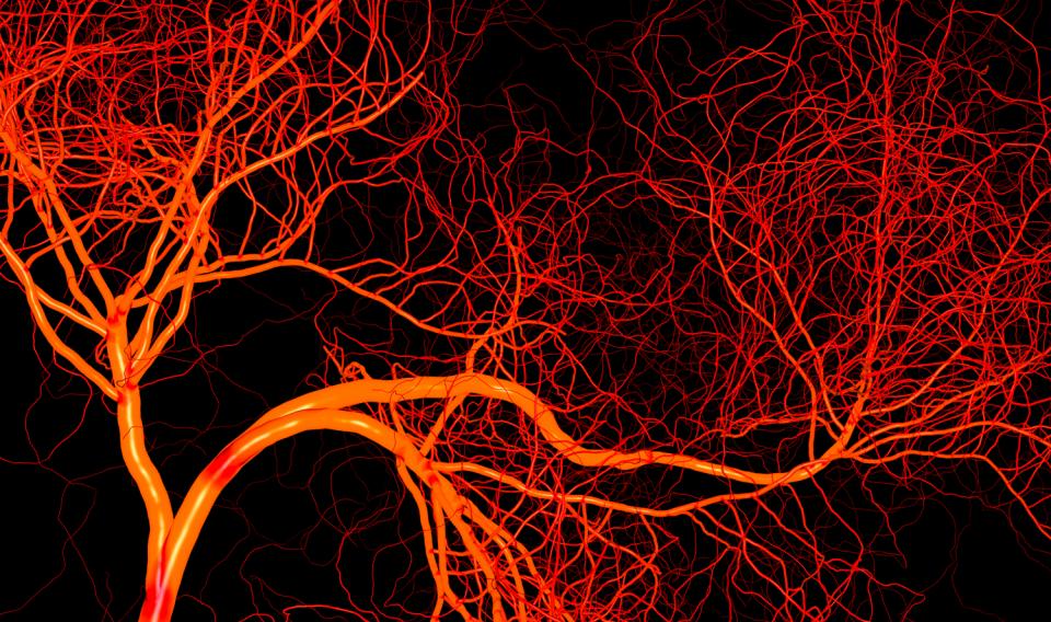 Network of vessels
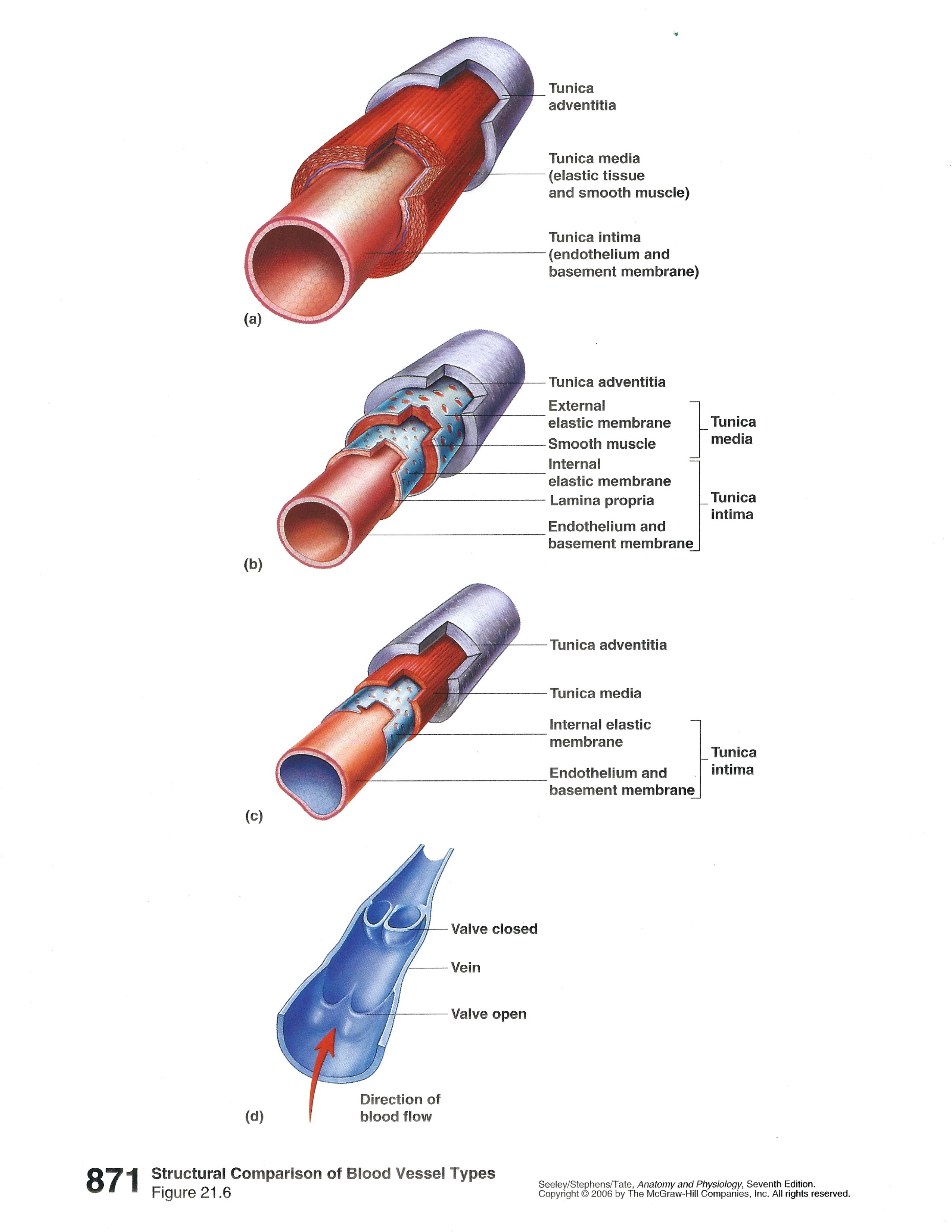
All blood vessels have one or ore layers around them depending on the type of of vessel, artery, arteriole, capillary, venue, or vein.
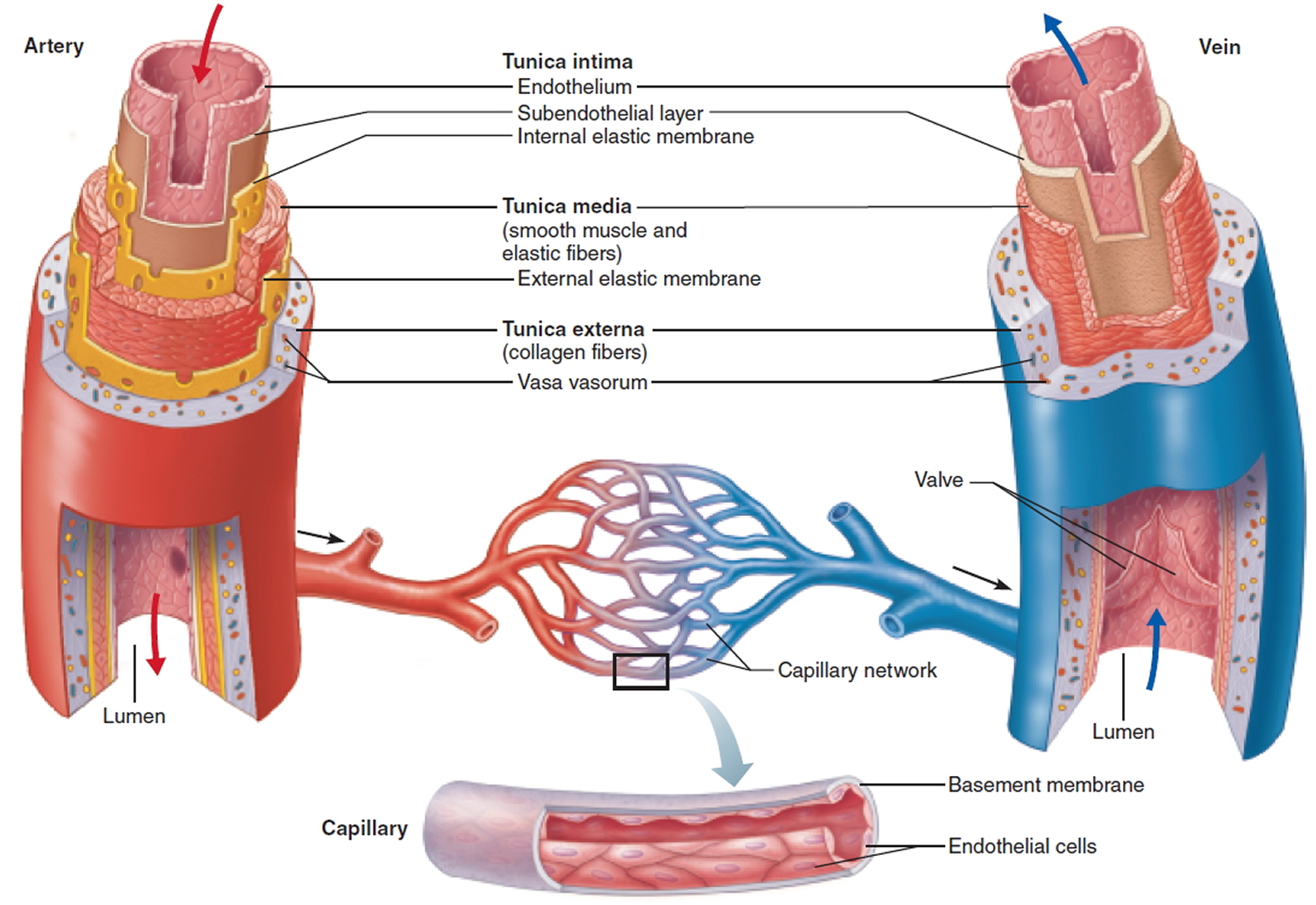
As you can see, there are considerable differences in the number and thickness of these layers. As usual structure is related to function. Arteries have to withstand the highest pressures as the heart contracts and forces blood into the system. that why you found elastic connective tissue in the walls of the aorta back in histology. Capillaries have a single, simple squamous layer to allow maximum diffusion of materials into or out of the vessel.
 Blood Pressure Graph
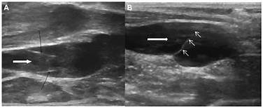 Image of Valves
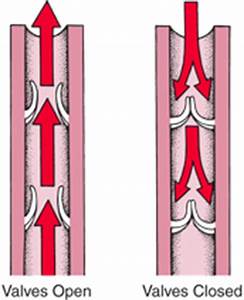
Veins have the lowest pressure and need valves to ensure blood flows in the correct direction in the vessel (just like lymphatic vessel). As you can see from the graph, blood pressure and velocity drop rapidly as it enters the capillaries because the total vessel volume rapidly increases.This provides maximum time for exchange of materials between vessels and cells.
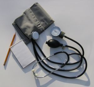
Blood pressure is a strong indicator of potential cardiovascular disease. It also tends to increase as you age--vessels are less flexible (arteriosclerosis) or may have fatty deposits in areas along the vessel walls (atherosclerosis). It is typically given as two numbers that represent systole in the heart and diastole in the heart--maximum pressure and lowest (or continuous) pressure. There are a number of devices for measuring but in many doctor's office the tried and true sphygmomanometer is still used.
This website provides more details. Structure and Function of Blood Vessels