Nervous System Organization
Nervous System Organization
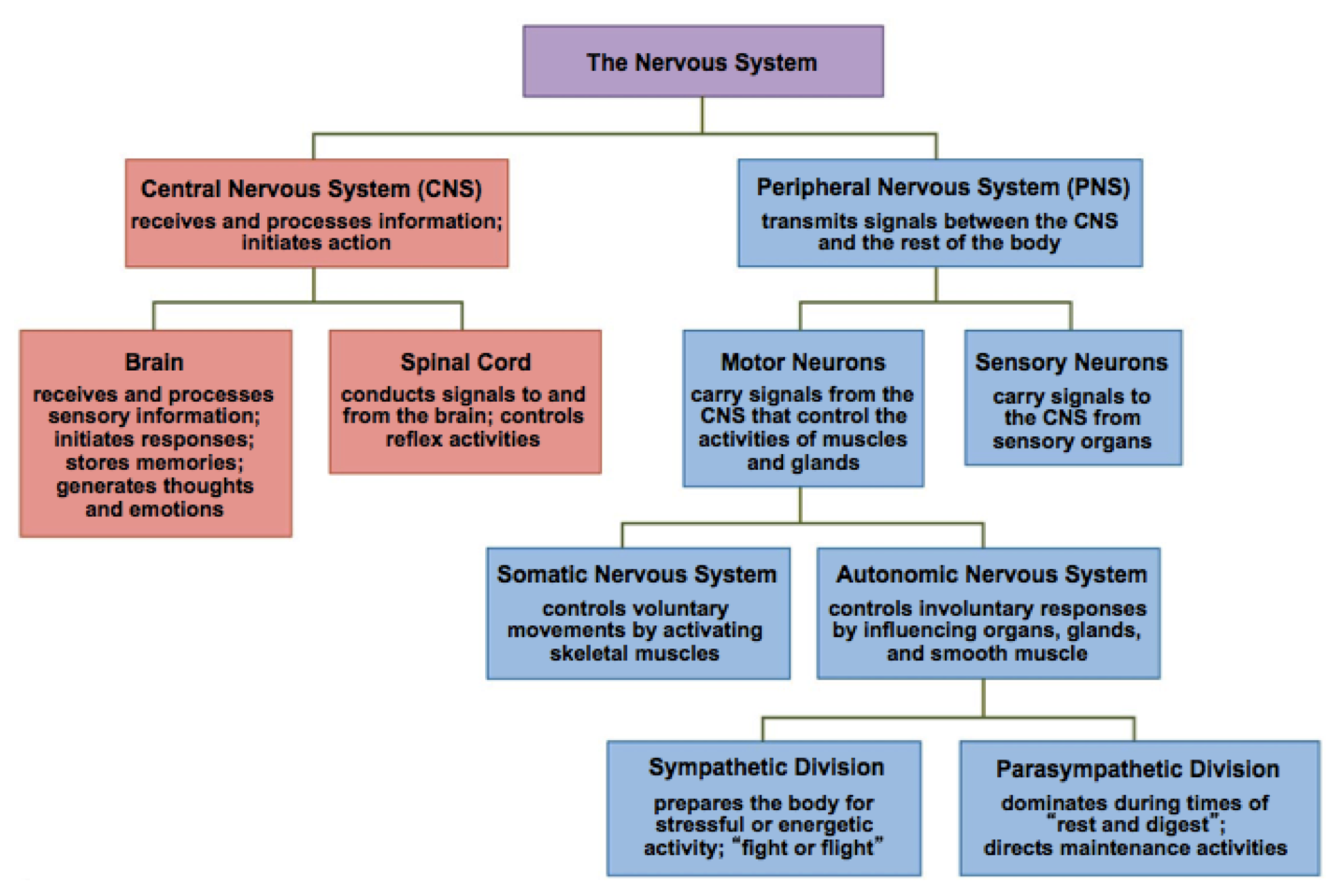
Vertebrate systems are generally divided into an information transmission portion (Peripheral Nervous System or PNS) and a central integration and decision portion (Central Nervous System or CNS).
The PNS is normally organized into a sensory system that carries information from the various sense organs to the CNS and a motor system that carries responses back to effecters. The motor system is further divided into a somatic system that was voluntary and controlled skeletal muscles and an autonomic system that is involuntary and controls internal organs and smooth muscle. The somatic subdivision carries information to skin, tendons, and muscles and is the voluntary portion. The autonomic subdivision carries information to the internal organs, glands, etc. It is generally considered to be involuntary. These are examined more closely in the text and in the site The Autonomic Nervous System (Links to an external site.).
Autonomic Nervous System
The Peripheral Nervous System is normally organized into a sensory system that carries information from the various sense organs to the CNS and a motor system that carries responses back to effecters. The motor system is further divided into a somatic system that was voluntary and controlled skeletal muscles and an autonomic system that is involuntary and controls internal organs and smooth muscle. The somatic subdivision carries information to skin, tendons, and muscles and is the voluntary portion. The autonomic subdivision carries information to the internal organs, glands, etc. It is generally considered to be involuntary. This section focuses on the autonomic system.
The autonomic system is in turn subdivided into two sections--the Sympathetic Nervous System and the Parasympathetic Nervous System. Structurally, each system is different. All sympathetic nerves originate from autonomic ganglia near the thoracic and lumbar portions of the spinal cord. Each nerve then extends to a particular organ or gland. The parasympathetic nerves originate from only two locations. Three cranial nerves originate from the brain and one of them, the vagus nerve, extends into the abdomen and connects to numerous internal organs. The other location is from the sacral portion of the spinal cord. Functionally, each of these systems enervates the same organs but have opposite effects on that organ or gland.
When the sympathetic system acts as a unit, it is part of the fight or flight response, the body’s response to what is perceived to be a stress or emergency situation. Under these conditions, the pupils dilate, bronchi and air passages in lungs dilate, salivary glands slow secretions, heart rate increases, digestive organs (stomach, intestines, pancreas) slow down, sphincters contract, adrenal medulla increases secretion of epinephrine (see text for more details). The parasympathetic has opposite effects from those above. The two systems maintain homeostatic conditions.
The CNS has two parts--the brain and the spinal cord. The spinal cord is primarily apathway carrying information to and from the brain from the different levels of the body. It has paired spinal nerves that leave between the vertebrae; motor and sensory nerves have different pathways as the enter and leave spinal cord but then come together to form the nerves going to and from different parts of the body. More detail on the spinal cord structure is at the link The Spinal Cord (Links to an external site.). Damage to the spinal cord generally does not heal. Much work is being done to determine if that can be changed (see Spinal Cord Repair (Links to an external site.)).
The final portion of the nervous system is the brain. The vertebrate brain consists of three primary areas as determined from embryonic development--the hindbrain, midbrain and forebrain. More detail is available at the link Divisions of the Brain (Links to an external site.). The reticular formation that later forms actually extends through all three portions of the brain.The brain and spinal cord are separated from most other body cells by a layer of glial cells forming the Blood-Brain Barrier (Links to an external site.). The brain and spinal cord originally form as a hollow tube. The remains of that opening in the brain are called ventricles in the adult brain and are filled with a fluid called the cerebral spinal fluid. See more detail at The Ventriclular System and CSF
Regions of the Vertebrate Brain
The final portion of the nervous system is the brain. The vertebrate brain consists of three primary areas as determined from embryonic development--the hindbrain, midbrain and forebrain. The reticular formation that later forms actually extends through all three portions of the brain. The brain and spinal cord are separated from most other body cells by a layer of glial cells forming the blood-brain barrier. The brain and spinal cord originally form as a hollow tube. The remains of that opening in the brain are called ventricles in the adult brain and are filled with a fluid called the cerebral spinal fluid. The brain is the single most complex organ known. While our knowledge is growing rapidly, we have only begun to do more than map the surface. Each of the three embryonic areas is further subdivided. Each part of the brain has unique structures, functions, and interacts with numerous other brain structures and structures outside the brain.
HINDBRAIN
Medulla Oblongata: The medulla is the widened area just inside the skull where the spinal cord joins the brain. It is often referred to (along with the rest of the hindbrain) as the "primitive" brain. This part of the brain controls a number of essential reflex actions required to maintain life. It regulates heart rate, blood pressure, swallowing, vomiting, coughing, and breathing rate. Thus damage to this part of the brain is usually fatal.
Pons: The pons also has centers for breathing rate. In addition, it regulates the flow of signals coming up the spinal cord to higher brain levels; some of the nerve tracts cross over to the opposite side of the brain at this location. It also regulates signals to and from the cerebellum.
Cerebellum: The cerebellum coordinates muscle coordination and body position information. When you learn a new task, you build pathways/connections within the cerebellum so that you don’t need to consciously control the movement of each muscle involved but merely need to do the activity. Walking is a prime example: walking requires the coordination of many muscles to both move and avoid falling down (loss of balance). Once you learn how to walk, you do that without conscious thought. There is also some recent evidence of cerebellum activity during memory formation.
MIDBRAIN
The midbrain is a small connecting region between the hindbrain and forebrain. It contains several internal structures. These structures are involved in visual and auditory reflexes. The area also relays information to and from the cerebral cortex. As part of the reticular formation, it filters sensory inputs and routes them to the correct part of the cerebral cortex. Thus, it helps regulate awareness and sleep—it effectively screens most sensory information when asleep and allows all or most information through when your awareness is high. The transition from wakefulness to sleep and vice versa is controlled by this area. All information on muscle tone and movements must pass through the area between cerebellum and the motor control areas of the cerebrum.
FOREBRAIN
Thalamus: The thalamus sorts out sensory input going to cerebrum and relays signals from cerebrum to lower portions of the brain.
Limbic System: Actually runs from midbrain on up into the forebrain. It governs emotions and is involved with behaviors that are self-gratifying as well as fearful. Signals from olfactory and auditory senses also come into the limbic system and cause the feelings we have when we detect a scent or a particular sound from our past. It has four regions.
Hypothalamus: The hypothalamus is specialized for regulation of homeostasis of body systems. In this capacity, it regulates behaviors related to hunger, thirst, sleep/wake cycles, water balance, temperature, and sexual activity. It also regulates behaviors associated with other emotions—sweating, vomiting.
Amygdala: The amygdala helps with socialization and pleasure/punishment associated with behaviors. Sexual arousal is also a part of this structure.
Hippocampus: Involved during strong emotions such as anger and sexual activity. It has also been recently shown to play a role in long-term memory formation.
Cingulate Nucleus: Affects motivation and risk taking behavior.
Olfactory Lobes: These lobes are very pronounced in comparison to rest of cerebral cortex in animals that rely greatly on their sense of smell for survival. In humans, they are relatively small.
Cerebrum: The large cerebral cortex characterizes mammals in general and primates in particular. The cerebrum is divided into two hemispheres that are each composed of four lobes. Each lobe has specific functions that are mirrored in the other hemisphere. In addition, each hemisphere has unique functions that are either not found or are much reduced in the other hemisphere. It is where all sensory inputs are interpreted (they are all just action potentials until then) and all motor activity initiated.
Occipital Lobe: The occipital lobe is primarily concerned with vision. There is a primary visual area where action potentials from the opposite side visual field (if left occipital lobe, right visual field) are received after passing through the thalamus. There is also a visual association area where the inputs are associated with past learned visual information.
Temporal Lobe: The temporal lobe is primarily concerned with auditory information and association. Here is where you determine if the sounds you are hearing are speech, music, or simply static noise. Since spoken language is auditory, an area of this lobe (Wernicke's area) also “translates” speech into "thoughts". Damage to this area results in an inability to understand speech.
Parietal Lobe: The parietal lobe is primarily a sensory receiving and association area. These sensations may be visceral (internal, such as when you feel an upset stomach) or from somatic (external--eyes, touch, etc) senses. In the primary somatosensory area, each body part has a specific section of the cortex where it is received. The more sensitive an area is, the larger the surface area dedicated to that body part.
Frontal Lobe: The frontal lobe has several functions. First, it is the primary motor area. Aligned with each sensory area in the parietal lobe is a motor area in the frontal lobe corresponding to that area. This includes the motor activity involved with speech (Broca's area) and motor activity involved with scanning motions of the eyes.
Corpus Callosum: The corpus callosum is the connection between the two cerebral hemispheres. Since not all activities are mirrored in the other part of the cortex, the corpus callosum is how the two sides communicate with each other. Damage or deliberate cutting of this connection lead to some interesting sensory results (see your text for details).
Several parts of the brain are involved in both short and long term memory formation. It is thought that memories are stored primarily in the cerebral cortex. In addition, there is some evidence that each side of the brain has a few activities for which it is specialized. Popularly, there is the idea that the right brain is cold and logical while the left brain is artistic and emotional. This is a gross oversimplification and it is not strictly sex related either. If you find this topic interesting, take the time to research the topic and get accurate information.
The final portion of the nervous system is the brain. The vertebrate brain consists of three primary areas as determined from embryonic development--the hindbrain, midbrain and forebrain. More detail is available at the link Divisions of the Brain (Links to an external site.). The reticular formation that later forms actually extends through all three portions of the brain.The entire CNS is separated from the rest of the body cells by three membranes called meninges. The brain and spinal cord are separated from most other body cells by a layer of glial cells forming the Blood-Brain Barrier (Links to an external site.). There are several types of glial cells that support CNS function. The brain and spinal cord originally form as a hollow tube. The remains of that opening in the brain are called ventricles in the adult brain and are filled with a fluid called the cerebral spinal fluid. See more detail at The Ventriclular System and CSF (Links to an external site.).
The brain is the single most complex organ known. While our knowledge is growing rapidly, we have only begun to do more than map the surface. Each of the three embryonic areas is further subdivided. Each part of the brain has both unique functions and interacts with numerous other brain structures and structures outside the brain. Start by reviewing the parts of the brain found in the forebrain, midbrain, and hindbrain at the Divisions of the Brain (Links to an external site.) link (ignore the "___encephalon" names--just look at the three major divisions). The Secret Life Of the Brain (Links to an external site.) is a highly interactive PBS site on the brain. Be sure to look 3D Brain Anatomy shockwave presentation and explore the brain by structure and explore Scanning the Brain to see how we learn more about its function. Look at the two pages Lobes of the Brain (Links to an external site.) and Brain Right Down the Middle (Links to an external site.) to see the structures of the brain and their functions. If you are interested in the right/left brain topic, look at One Brain or Two? (Links to an external site.) and Splitting the Human Brain (Links to an external site.).There is also an excellent 3-D version of the brain at the site Genes to Cognition
Finally, be sure to review how memories are formed (Links to an external site.) and Human Memory Processing (Links to an external site.), how the brain "matures", and how drugs affect the brain (Links to an external site--scroll down TOC page..) and other parts of the nervous system.